 Movie
Movie Controller

[English] 日本語
 Yorodumi
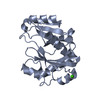
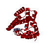
- PDB-6zpb: Cyanophage S-2L HD phosphohydrolase (DatZ) bound to dA and two ca... -
+ Open data

- Basic information
| Entry | Database: PDB / ID: 6zpb | ||||||
|---|---|---|---|---|---|---|---|
| Title | Cyanophage S-2L HD phosphohydrolase (DatZ) bound to dA and two catalytic Co2+ ions | ||||||
 Components Components | DatZ | ||||||
 Keywords Keywords | VIRAL PROTEIN / S-2L / HD phosphohydrolase / DatZ | ||||||
| Function / homology | Chem-3D1 / :  Function and homology information Function and homology information | ||||||
| Biological species |  Cyanophage S-2L (virus) Cyanophage S-2L (virus) | ||||||
| Method |  X-RAY DIFFRACTION / SYNCHROTRON / SAD / Resolution: 1.72097384171 Å X-RAY DIFFRACTION / SYNCHROTRON / SAD / Resolution: 1.72097384171 Å | ||||||
 Authors Authors | Czernecki, D. / Legrand, P. / Delarue, M. | ||||||
 Citation Citation | Journal: Nat Commun / Year: 2021 Title: How cyanophage S-2L rejects adenine and incorporates 2-aminoadenine to saturate hydrogen bonding in its DNA. Authors: Czernecki, D. / Legrand, P. / Tekpinar, M. / Rosario, S. / Kaminski, P.A. / Delarue, M. | ||||||
| History |
|
- Structure visualization
| Structure viewer | Molecule: MolmilJmol/JSmol |
|---|
- Downloads & links
-Download
| PDBx/mmCIF format | 6zpb.cif.gz | 63.5 KB | Display | PDBx/mmCIF format |
|---|---|---|---|---|
| PDB format | pdb6zpb.ent.gz | 40.8 KB | Display | PDB format |
| PDBx/mmJSON format | 6zpb.json.gz | Tree view | PDBx/mmJSON format | |
| Others |  Other downloads Other downloads |
-Validation report
| Arichive directory | https://data.pdbj.org/pub/pdb/validation_reports/zp/6zpbftp://data.pdbj.org/pub/pdb/validation_reports/zp/6zpb | HTTPS FTP |
|---|
-Related structure data


-Links
 PDBj
PDBj
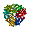
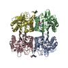
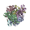
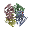
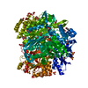
- Assembly
| Deposited unit | 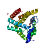
| |||||||||||||||||||||||||||
|---|---|---|---|---|---|---|---|---|---|---|---|---|---|---|---|---|---|---|---|---|---|---|---|---|---|---|---|---|
| 1 | x 6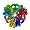
| |||||||||||||||||||||||||||
| Unit cell |
| |||||||||||||||||||||||||||
| Components on special symmetry positions |
|
-Components
| #1: Protein | Mass: 20612.609 Da / Num. of mol.: 1 Source method: isolated from a genetically manipulated source Source: (gene. exp.) Cyanophage S-2L (virus) / Production host:  | ||||
|---|---|---|---|---|---|
| #2: Chemical | ChemComp-3D1 / (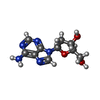  Mass: 251.242 Da / Num. of mol.: 1 / Source method: obtained synthetically / Formula: C10H13N5O3 / Feature type: SUBJECT OF INVESTIGATION Mass: 251.242 Da / Num. of mol.: 1 / Source method: obtained synthetically / Formula: C10H13N5O3 / Feature type: SUBJECT OF INVESTIGATION | ||||
| #3: Chemical |   Mass: 58.933 Da / Num. of mol.: 3 / Source method: obtained synthetically / Formula: Co / Feature type: SUBJECT OF INVESTIGATION Mass: 58.933 Da / Num. of mol.: 3 / Source method: obtained synthetically / Formula: Co / Feature type: SUBJECT OF INVESTIGATION#4: Water | ChemComp-HOH / |  Mass: 18.015 Da / Num. of mol.: 260 / Source method: isolated from a natural source / Formula: H2O Mass: 18.015 Da / Num. of mol.: 260 / Source method: isolated from a natural source / Formula: H2OHas ligand of interest | Y | |
-Experimental details
-Experiment
| Experiment | Method: X-RAY DIFFRACTION / Number of used crystals: 1 |
|---|
- Sample preparation
| Crystal | Density Matthews: 2.53 Å3/Da / Density % sol: 51.29 % |
|---|---|
| Crystal grow | Temperature: 291.15 K / Method: vapor diffusion, hanging drop / Details: 1.5 M LiSO4; 100 mM HEPES; 10 mM CoCl2 |
-Data collection
| Diffraction | Mean temperature: 100 K / Serial crystal experiment: N |
|---|---|
| Diffraction source | Source: SYNCHROTRON / Site: SOLEIL  / Beamline: PROXIMA 1 / Wavelength: 1.033202 Å / Beamline: PROXIMA 1 / Wavelength: 1.033202 Å |
| Detector | Type: DECTRIS EIGER X 16M / Detector: PIXEL / Date: Dec 7, 2018 / Details: KB Mirrors |
| Radiation | Monochromator: Si 111 / Protocol: SINGLE WAVELENGTH / Monochromatic (M) / Laue (L): M / Scattering type: x-ray |
| Radiation wavelength | Wavelength: 1.033202 Å / Relative weight: 1 |
| Reflection | Resolution: 1.72→40.44 Å / Num. obs: 21613 / % possible obs: 98.4 % / Redundancy: 14.2 % / Biso Wilson estimate: 14.0930917762 Å2 / CC1/2: 0.999 / Rmerge(I) obs: 0.083 / Rpim(I) all: 0.022 / Rrim(I) all: 0.086 / Net I/σ(I): 25 |
| Reflection shell | Resolution: 1.72→1.77 Å / Redundancy: 5.7 % / Rmerge(I) obs: 0.307 / Mean I/σ(I) obs: 5.1 / Num. unique obs: 1400 / CC1/2: 0.939 / Rpim(I) all: 0.137 / Rrim(I) all: 0.338 / % possible all: 88 |
- Processing
| Software |
| |||||||||||||||||||||||||||||||||||||||||||||||||||||||||||||||
|---|---|---|---|---|---|---|---|---|---|---|---|---|---|---|---|---|---|---|---|---|---|---|---|---|---|---|---|---|---|---|---|---|---|---|---|---|---|---|---|---|---|---|---|---|---|---|---|---|---|---|---|---|---|---|---|---|---|---|---|---|---|---|---|---|
| Refinement | Method to determine structure: SAD / Resolution: 1.72097384171→40.4358636633 Å / SU ML: 0.165812802632 / Cross valid method: FREE R-VALUE / σ(F): 1.45045910224 / Phase error: 16.9899637983 Stereochemistry target values: GeoStd + Monomer Library + CDL v1.2
| |||||||||||||||||||||||||||||||||||||||||||||||||||||||||||||||
| Solvent computation | Shrinkage radii: 0.9 Å / VDW probe radii: 1.11 Å / Solvent model: FLAT BULK SOLVENT MODEL | |||||||||||||||||||||||||||||||||||||||||||||||||||||||||||||||
| Displacement parameters | Biso mean: 17.5063621561 Å2 | |||||||||||||||||||||||||||||||||||||||||||||||||||||||||||||||
| Refinement step | Cycle: LAST / Resolution: 1.72097384171→40.4358636633 Å
| |||||||||||||||||||||||||||||||||||||||||||||||||||||||||||||||
| Refine LS restraints |
| |||||||||||||||||||||||||||||||||||||||||||||||||||||||||||||||
| LS refinement shell |
|
