 Movie
Movie Controller


+ Open data

- Basic information
| Entry | Database: PDB / ID: 6zmb | ||||||
|---|---|---|---|---|---|---|---|
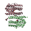
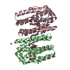
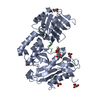
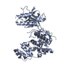
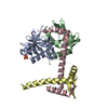
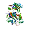
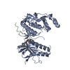
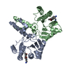
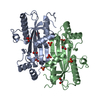
| Title | Structure of the native tRNA-Monooxygenase enzyme MiaE | ||||||
 Components Components | tRNA hydroxylase | ||||||
 Keywords Keywords | OXIDOREDUCTASE / tRNA-monooxygenase metallo-enzyme tRNA-modifying enzyme hydroxylase | ||||||
| Function / homology |  Function and homology information Function and homology informationtRNA 2-(methylsulfanyl)-N(6)-isopentenyladenosine(37) hydroxylase activity / tRNA modification / metal ion binding Similarity search - Function | ||||||
| Biological species |  Pseudomonas putida KT2440 (bacteria) Pseudomonas putida KT2440 (bacteria) | ||||||
| Method |  X-RAY DIFFRACTION / SYNCHROTRON / MOLECULAR REPLACEMENT / Resolution: 1.7 Å X-RAY DIFFRACTION / SYNCHROTRON / MOLECULAR REPLACEMENT / Resolution: 1.7 Å | ||||||
 Authors Authors | Carpentier, P. / Atta, M. | ||||||
| Funding support |  France, 1items France, 1items
| ||||||
 Citation Citation | Journal: Nucleic Acids Res. / Year: 2020 Title: Structural, biochemical and functional analyses of tRNA-monooxygenase enzyme MiaE from Pseudomonas putida provide insights into tRNA/MiaE interaction. Authors: Carpentier, P. / Lepretre, C. / Basset, C. / Douki, T. / Torelli, S. / Duarte, V. / Hamdane, D. / Fontecave, M. / Atta, M. | ||||||
| History |
|
- Structure visualization
| Structure viewer | Molecule: MolmilJmol/JSmol |
|---|
- Downloads & links
-Download
| PDBx/mmCIF format | 6zmb.cif.gz | 105.7 KB | Display | PDBx/mmCIF format |
|---|---|---|---|---|
| PDB format | pdb6zmb.ent.gz | 79.2 KB | Display | PDB format |
| PDBx/mmJSON format | 6zmb.json.gz | Tree view | PDBx/mmJSON format | |
| Others |  Other downloads Other downloads |
-Validation report
| Arichive directory | https://data.pdbj.org/pub/pdb/validation_reports/zm/6zmbftp://data.pdbj.org/pub/pdb/validation_reports/zm/6zmb | HTTPS FTP |
|---|
-Related structure data
| Related structure data |  6zmaC  6zmcC 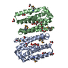 2itbS S: Starting model for refinement C: citing same article ( |
|---|---|
| Similar structure data |

-Links
 PDBj
PDBj


- Assembly
| Deposited unit | 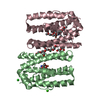
| ||||||||||||||||||
|---|---|---|---|---|---|---|---|---|---|---|---|---|---|---|---|---|---|---|---|
| 1 |
| ||||||||||||||||||
| Unit cell |
| ||||||||||||||||||
| Components on special symmetry positions |
|
-Components
-Protein , 1 types, 2 molecules BC
| #1: Protein | Mass: 22661.078 Da / Num. of mol.: 2 Source method: isolated from a genetically manipulated source Source: (gene. exp.) Pseudomonas putida KT2440 (bacteria) / Gene: AYO08_21560, CBL13_00280, CBP06_18695 / Production host: |
|---|
-Non-polymers , 9 types, 232 molecules 


| #2: Chemical | ChemComp-FE /  Mass: 55.845 Da / Num. of mol.: 4 / Source method: obtained synthetically / Formula: Fe / Feature type: SUBJECT OF INVESTIGATION Mass: 55.845 Da / Num. of mol.: 4 / Source method: obtained synthetically / Formula: Fe / Feature type: SUBJECT OF INVESTIGATION#3: Chemical |  Mass: 35.453 Da / Num. of mol.: 2 / Source method: obtained synthetically / Formula: Cl Mass: 35.453 Da / Num. of mol.: 2 / Source method: obtained synthetically / Formula: Cl#4: Chemical | ChemComp-CA / |  Mass: 40.078 Da / Num. of mol.: 1 / Source method: obtained synthetically / Formula: Ca Mass: 40.078 Da / Num. of mol.: 1 / Source method: obtained synthetically / Formula: Ca#5: Chemical |  Mass: 122.143 Da / Num. of mol.: 2 / Source method: obtained synthetically / Formula: C4H12NO3 / Comment: pH buffer*YM Mass: 122.143 Da / Num. of mol.: 2 / Source method: obtained synthetically / Formula: C4H12NO3 / Comment: pH buffer*YM#6: Chemical |  Mass: 106.120 Da / Num. of mol.: 2 / Source method: obtained synthetically / Formula: C4H10O3 Mass: 106.120 Da / Num. of mol.: 2 / Source method: obtained synthetically / Formula: C4H10O3#7: Chemical | ChemComp-PG4 / |  Mass: 194.226 Da / Num. of mol.: 1 / Source method: obtained synthetically / Formula: C8H18O5 / Comment: precipitant*YM Mass: 194.226 Da / Num. of mol.: 1 / Source method: obtained synthetically / Formula: C8H18O5 / Comment: precipitant*YM#8: Chemical |  Mass: 150.173 Da / Num. of mol.: 2 / Source method: obtained synthetically / Formula: C6H14O4 Mass: 150.173 Da / Num. of mol.: 2 / Source method: obtained synthetically / Formula: C6H14O4#9: Chemical | ChemComp-PG6 / |  Mass: 266.331 Da / Num. of mol.: 1 / Source method: obtained synthetically / Formula: C12H26O6 Mass: 266.331 Da / Num. of mol.: 1 / Source method: obtained synthetically / Formula: C12H26O6#10: Water | ChemComp-HOH / | Mass: 18.015 Da / Num. of mol.: 217 / Source method: isolated from a natural source / Formula: H2O |
|---|
-Details
| Has ligand of interest | Y |
|---|
-Experimental details
-Experiment
| Experiment | Method: X-RAY DIFFRACTION / Number of used crystals: 1 |
|---|
- Sample preparation
| Crystal | Density Matthews: 2.51 Å3/Da / Density % sol: 50.93 % / Description: elongated |
|---|---|
| Crystal grow | Temperature: 294 K / Method: vapor diffusion, hanging drop / pH: 8 Details: MiaE: 20 mg/ml in 100 mM HEPES, pH 7.5, 30mM NaCl reservoir solution: 0.5M CaCl2, 42% PEG 6k, 2M Tris-Cl pH 8 |
-Data collection
| Diffraction | Mean temperature: 100 K / Serial crystal experiment: N |
|---|---|
| Diffraction source | Source: SYNCHROTRON / Site: ESRF / Beamline: ID29 / Wavelength: 0.9753 Å |
| Detector | Type: DECTRIS PILATUS 6M / Detector: PIXEL / Date: Jan 29, 2018 |
| Radiation | Protocol: SINGLE WAVELENGTH / Monochromatic (M) / Laue (L): M / Scattering type: x-ray |
| Radiation wavelength | Wavelength: 0.9753 Å / Relative weight: 1 |
| Reflection | Resolution: 1.7→50 Å / Num. obs: 48290 / % possible obs: 97.5 % / Observed criterion σ(I): 1.44 / Redundancy: 2.8 % / Biso Wilson estimate: 38.8 Å2 / CC1/2: 0.999 / Rrim(I) all: 0.04 / Net I/σ(I): 13.1 |
| Reflection shell | Resolution: 1.7→1.8 Å / Redundancy: 2.8 % / Num. unique obs: 7733 / CC1/2: 0.845 / Rrim(I) all: 0.846 / % possible all: 97.7 |
- Processing
| Software |
| ||||||||||||||||||||||||
|---|---|---|---|---|---|---|---|---|---|---|---|---|---|---|---|---|---|---|---|---|---|---|---|---|---|
| Refinement | Method to determine structure: MOLECULAR REPLACEMENT Starting model: 2ITB Resolution: 1.7→46.752 Å / SU ML: 0.25 / Cross valid method: THROUGHOUT / σ(F): 1.33 / Phase error: 26.89 / Stereochemistry target values: ML
| ||||||||||||||||||||||||
| Solvent computation | Shrinkage radii: 0.9 Å / VDW probe radii: 1.11 Å / Solvent model: FLAT BULK SOLVENT MODEL | ||||||||||||||||||||||||
| Displacement parameters | Biso max: 92.64 Å2 / Biso mean: 36.955 Å2 / Biso min: 18.88 Å2 | ||||||||||||||||||||||||
| Refinement step | Cycle: final / Resolution: 1.7→46.752 Å
| ||||||||||||||||||||||||
| LS refinement shell | Resolution: 1.7→1.73 Å /
|
