 Movie
Movie Controller


+ Open data

- Basic information
| Entry | Database: PDB / ID: 6zjk | ||||||
|---|---|---|---|---|---|---|---|
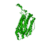
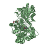
| Title | Ribonucleotide reductase R2 subunit from Clostridium botulinum | ||||||
 Components Components | Ribonucleoside-diphosphate reductase subunit beta | ||||||
 Keywords Keywords | METAL BINDING PROTEIN / ribonucleotide / reductase / nucleotide / R2 | ||||||
| Function / homology |  Function and homology information Function and homology informationribonucleoside-diphosphate reductase / ribonucleoside-diphosphate reductase activity, thioredoxin disulfide as acceptor / deoxyribonucleotide biosynthetic process / metal ion binding Similarity search - Function | ||||||
| Biological species |   Clostridium botulinum (bacteria) Clostridium botulinum (bacteria) | ||||||
| Method |  X-RAY DIFFRACTION / SYNCHROTRON / MOLECULAR REPLACEMENT / Resolution: 2 Å X-RAY DIFFRACTION / SYNCHROTRON / MOLECULAR REPLACEMENT / Resolution: 2 Å | ||||||
 Authors Authors | Martinez-Carranza, M. / Stenmark, P. | ||||||
 Citation Citation | Journal: J.Biol.Chem. / Year: 2020 Title: A ribonucleotide reductase from Clostridium botulinum reveals distinct evolutionary pathways to regulation via the overall activity site. Authors: Martinez-Carranza, M. / Jonna, V.R. / Lundin, D. / Sahlin, M. / Carlson, L.A. / Jemal, N. / Hogbom, M. / Sjoberg, B.M. / Stenmark, P. / Hofer, A. | ||||||
| History |
|
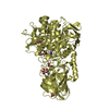
- Structure visualization
| Structure viewer | Molecule: MolmilJmol/JSmol |
|---|
- Downloads & links
-Download
| PDBx/mmCIF format | 6zjk.cif.gz | 543.7 KB | Display | PDBx/mmCIF format |
|---|---|---|---|---|
| PDB format | pdb6zjk.ent.gz | 446.6 KB | Display | PDB format |
| PDBx/mmJSON format | 6zjk.json.gz | Tree view | PDBx/mmJSON format | |
| Others |  Other downloads Other downloads |
-Validation report
| Summary document | 6zjk_validation.pdf.gz | 4.9 MB | Display | wwPDB validaton report |
|---|---|---|---|---|
| Full document | 6zjk_full_validation.pdf.gz | 4.9 MB | Display | |
| Data in XML | 6zjk_validation.xml.gz | 46.4 KB | Display | |
| Data in CIF | 6zjk_validation.cif.gz | 65.2 KB | Display | |
| Arichive directory | https://data.pdbj.org/pub/pdb/validation_reports/zj/6zjkftp://data.pdbj.org/pub/pdb/validation_reports/zj/6zjk | HTTPS FTP |
-Related structure data
| Related structure data |  2rccS S: Starting model for refinement |
|---|---|
| Similar structure data |


-Links
 PDBj
PDBj
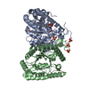
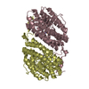
- Assembly
| Deposited unit | 
| ||||||||
|---|---|---|---|---|---|---|---|---|---|
| 1 | 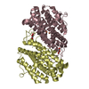
| ||||||||
| 2 | 
| ||||||||
| Unit cell |
| ||||||||
| Components on special symmetry positions |
|
-Components
| #1: Protein | Mass: 43246.566 Da / Num. of mol.: 4 Source method: isolated from a genetically manipulated source Source: (gene. exp.) Clostridium botulinum (strain Loch Maree / Type A3) (bacteria)Strain: Loch Maree / Type A3 / Gene: nrdB, CLK_2200 / Production host: References: UniProt: B1KYY8, ribonucleoside-diphosphate reductase #2: Chemical | ChemComp-GOL /   Mass: 92.094 Da / Num. of mol.: 4 / Source method: obtained synthetically / Formula: C3H8O3 Mass: 92.094 Da / Num. of mol.: 4 / Source method: obtained synthetically / Formula: C3H8O3#3: Chemical | ChemComp-FE /   Mass: 55.845 Da / Num. of mol.: 8 / Source method: obtained synthetically / Formula: Fe / Feature type: SUBJECT OF INVESTIGATION Mass: 55.845 Da / Num. of mol.: 8 / Source method: obtained synthetically / Formula: Fe / Feature type: SUBJECT OF INVESTIGATION#4: Water | ChemComp-HOH / |  Mass: 18.015 Da / Num. of mol.: 193 / Source method: isolated from a natural source / Formula: H2O Mass: 18.015 Da / Num. of mol.: 193 / Source method: isolated from a natural source / Formula: H2OHas ligand of interest | Y | |
|---|
-Experimental details
-Experiment
| Experiment | Method: X-RAY DIFFRACTION / Number of used crystals: 1 |
|---|
- Sample preparation
| Crystal | Density Matthews: 2.46 Å3/Da / Density % sol: 49.9 % |
|---|---|
| Crystal grow | Temperature: 294.15 K / Method: vapor diffusion, sitting drop / Details: sodium bromide, Bis Tris propane pH 6.5, PEG 3350 |
-Data collection
| Diffraction | Mean temperature: 115 K / Serial crystal experiment: N |
|---|---|
| Diffraction source | Source: SYNCHROTRON / Site: Diamond  / Beamline: I04-1 / Wavelength: 0.92 Å / Beamline: I04-1 / Wavelength: 0.92 Å |
| Detector | Type: DECTRIS PILATUS 6M-F / Detector: PIXEL / Date: Jul 1, 2016 |
| Radiation | Protocol: SINGLE WAVELENGTH / Monochromatic (M) / Laue (L): M / Scattering type: x-ray |
| Radiation wavelength | Wavelength: 0.92 Å / Relative weight: 1 |
| Reflection | Limit h max: 99 / Limit h min: -103 / Limit k max: 19 / Limit k min: -20 / Limit l max: 75 / Limit l min: -74 / Number: 123556 / Theta max: 13.345875285 ° / Theta min: 0.544372704633 ° |
| Reflection | Resolution: 2→83.68 Å / Num. obs: 106664 / % possible obs: 99.6 % / Redundancy: 6.6 % / Biso Wilson estimate: 39.35 Å2 / CC1/2: 0.999 / Rmerge(I) obs: 0.073 / Net I/σ(I): 10.71 |
| Reflection shell | Resolution: 2→2.05 Å / Num. unique obs: 7815 / CC1/2: 0.664 |
| Cell measurement | Reflection used: 123556 / Theta max: 13.345875285 ° / Theta min: 0.544372704633 ° |
- Processing
| Software |
| ||||||||||||||||||||||||||||||||||||||||||||||||
|---|---|---|---|---|---|---|---|---|---|---|---|---|---|---|---|---|---|---|---|---|---|---|---|---|---|---|---|---|---|---|---|---|---|---|---|---|---|---|---|---|---|---|---|---|---|---|---|---|---|
| Refinement | Method to determine structure: MOLECULAR REPLACEMENT Starting model: 2RCC Resolution: 2→83.67 Å / SU ML: 0.32 / Cross valid method: THROUGHOUT / σ(F): 1.34 / Phase error: 31.53 / Stereochemistry target values: ML
| ||||||||||||||||||||||||||||||||||||||||||||||||
| Solvent computation | Shrinkage radii: 0.9 Å / VDW probe radii: 1.11 Å / Solvent model: FLAT BULK SOLVENT MODEL | ||||||||||||||||||||||||||||||||||||||||||||||||
| Displacement parameters | Biso max: 114.08 Å2 / Biso mean: 54.6969 Å2 / Biso min: 31.96 Å2 | ||||||||||||||||||||||||||||||||||||||||||||||||
| Refinement step | Cycle: final / Resolution: 2→83.67 Å
| ||||||||||||||||||||||||||||||||||||||||||||||||
| Refinement TLS params. | Method: refined / Origin x: -7.0152 Å / Origin y: 15.2283 Å / Origin z: 32.2143 Å
| ||||||||||||||||||||||||||||||||||||||||||||||||
| Refinement TLS group |
|
