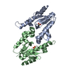
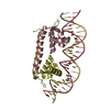
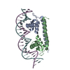
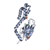
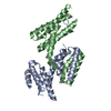
Regulation of HSF1-mediated heat shock response / mRNA Splicing - Major Pathway / HSP90 chaperone cycle for steroid hormone receptors (SHR) in the presence of ligand / AUF1 (hnRNP D0) binds and destabilizes mRNA / Neutrophil degranulation / heat shock protein binding / protein folding chaperone / ATP-dependent protein folding chaperone / protein refolding / ATP hydrolysis activity ...Regulation of HSF1-mediated heat shock response / mRNA Splicing - Major Pathway / HSP90 chaperone cycle for steroid hormone receptors (SHR) in the presence of ligand / AUF1 (hnRNP D0) binds and destabilizes mRNA / Neutrophil degranulation / heat shock protein binding / protein folding chaperone / ATP-dependent protein folding chaperone / protein refolding / ATP hydrolysis activity / ATP binding / metal ion binding / nucleus / cytoplasm Similarity search - Function
Substrate Binding Domain Of Dnak; Chain:A; Domain 2 - #10 / Substrate Binding Domain Of DNAk; Chain A, domain 1 / Substrate Binding Domain Of DNAk; Chain A, domain 1 / Substrate Binding Domain Of Dnak; Chain:A; Domain 2 / Heat shock hsp70 proteins family signature 2. / Heat shock hsp70 proteins family signature 1. / Heat shock hsp70 proteins family signature 3. / Heat shock protein 70, conserved site / Heat shock protein 70kD, peptide-binding domain superfamily / Heat shock protein 70kD, C-terminal domain superfamily ...Substrate Binding Domain Of Dnak; Chain:A; Domain 2 - #10 / Substrate Binding Domain Of DNAk; Chain A, domain 1 / Substrate Binding Domain Of DNAk; Chain A, domain 1 / Substrate Binding Domain Of Dnak; Chain:A; Domain 2 / Heat shock hsp70 proteins family signature 2. / Heat shock hsp70 proteins family signature 1. / Heat shock hsp70 proteins family signature 3. / Heat shock protein 70, conserved site / Heat shock protein 70kD, peptide-binding domain superfamily / Heat shock protein 70kD, C-terminal domain superfamily / Heat shock protein 70 family / Hsp70 protein / ATPase, nucleotide binding domain / Up-down Bundle / Sandwich / Mainly Beta / Mainly Alpha Similarity search - Domain/homology
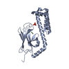
Mass: 25835.967 Da / Num. of mol.: 4 Source method: isolated from a genetically manipulated source Source: (gene. exp.) Plasmodium falciparum (isolate 3D7) (eukaryote) Gene: PF3D7_0831700 / Plasmid: pFLOAT Details (production host): pET30a derivative with His-tag and 3C cleavage site Production host: Escherichia coli BL21(DE3) (bacteria) / Variant (production host): Rosetta2 / References: UniProt: K7NTP5
#2: Protein/peptide
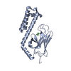
ASN-ARG-LEU-LEU-LEU-THR-GLY
Mass: 786.941 Da / Num. of mol.: 4 / Source method: obtained synthetically Source: (synth.) Plasmodium falciparum (malaria parasite P. falciparum)
-
Experimental details
-
Experiment
Experiment
Method: X-RAY DIFFRACTION / Number of used crystals: 1
-
Sample preparation
Crystal
Density Matthews: 2.8 Å3/Da / Density % sol: 56.04 %
Crystal grow
Temperature: 293 K / Method: vapor diffusion, sitting drop / pH: 8.5 Details: 0.1 M bicine/2-Amino-2-(hydroxymethyl)propane-1,3-diol base 10% w/v PEG 20,000 20% w/v PEG MME 550 0.03 M of each of di- to penta-ethyleneglycol
-
Data collection
Diffraction
Mean temperature: 100 K / Serial crystal experiment: N
In the structure databanks used in Yorodumi, some data are registered as the other names, "COVID-19 virus" and "2019-nCoV". Here are the details of the virus and the list of structure data.
Jan 31, 2019. EMDB accession codes are about to change! (news from PDBe EMDB page)
EMDB accession codes are about to change! (news from PDBe EMDB page)
The allocation of 4 digits for EMDB accession codes will soon come to an end. Whilst these codes will remain in use, new EMDB accession codes will include an additional digit and will expand incrementally as the available range of codes is exhausted. The current 4-digit format prefixed with “EMD-” (i.e. EMD-XXXX) will advance to a 5-digit format (i.e. EMD-XXXXX), and so on. It is currently estimated that the 4-digit codes will be depleted around Spring 2019, at which point the 5-digit format will come into force.
The EM Navigator/Yorodumi systems omit the EMD- prefix.
Related info.:Q: What is EMD? / ID/Accession-code notation in Yorodumi/EM Navigator
Yorodumi is a browser for structure data from EMDB, PDB, SASBDB, etc.
This page is also the successor to EM Navigator detail page, and also detail information page/front-end page for Omokage search.
The word "yorodu" (or yorozu) is an old Japanese word meaning "ten thousand". "mi" (miru) is to see.
Related info.:EMDB / PDB / SASBDB / Comparison of 3 databanks / Yorodumi Search / Aug 31, 2016. New EM Navigator & Yorodumi / Yorodumi Papers / Jmol/JSmol / Function and homology information / Changes in new EM Navigator and Yorodumi
 Movie
Movie Controller

 Yorodumi
 Open data
 Basic information
 Components
 Keywords
 Function and homology information
 X-RAY DIFFRACTION /
  Authors
 United Kingdom, 1items
  Citation
 Structure visualization
 Downloads & links
 Other downloads
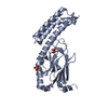


 PDBj
PDBj
 Assembly
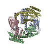


 Sample preparation
 Processing