 Movie
Movie Controller

[English] 日本語
 Yorodumi
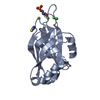
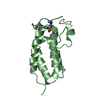
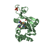
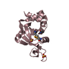
- PDB-6z96: [4Fe-4S]-dependent thiouracil desulfidase TudS (DUF523Vcz) soaked... -
+ Open data

- Basic information
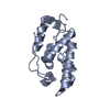
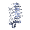
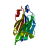
| Entry | Database: PDB / ID: 6z96 | ||||||
|---|---|---|---|---|---|---|---|
| Title | [4Fe-4S]-dependent thiouracil desulfidase TudS (DUF523Vcz) soaked with 4-thiouracil (12.65 keV, 7.125 keV (Fe-SAD) and 6.5 keV (S-SAD) data) | ||||||
 Components Components | TudS | ||||||
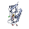
 Keywords Keywords | TRANSFERASE / thiouracil / iron-sulfur cluster / desulfuration / sulfurtransferase | ||||||
| Function / homology | 2-thiouracil desulfurase / 2-thiouracil desulfurase / 4 iron, 4 sulfur cluster binding / metal ion binding / HYDROSULFURIC ACID / DI(HYDROXYETHYL)ETHER / IRON/SULFUR CLUSTER / DUF523 domain-containing protein Function and homology information Function and homology information | ||||||
| Biological species |  uncultured bacterium (environmental samples) uncultured bacterium (environmental samples) | ||||||
| Method |  X-RAY DIFFRACTION / SYNCHROTRON / MOLECULAR REPLACEMENT / Resolution: 1.327 Å X-RAY DIFFRACTION / SYNCHROTRON / MOLECULAR REPLACEMENT / Resolution: 1.327 Å | ||||||
 Authors Authors | Pecqueur, L. / Zhou, J. / Fontecave, M. / Golinelli-Pimpaneau, B. | ||||||
| Funding support |  France, 1items France, 1items
| ||||||
 Citation Citation | Journal: Angew.Chem.Int.Ed.Engl. / Year: 2021 Title: Structural Evidence for a [4Fe-5S] Intermediate in the Non-Redox Desulfuration of Thiouracil. Authors: Zhou, J. / Pecqueur, L. / Aucynaite, A. / Fuchs, J. / Rutkiene, R. / Vaitekunas, J. / Meskys, R. / Boll, M. / Fontecave, M. / Urbonavicius, J. / Golinelli-Pimpaneau, B. | ||||||
| History |
|
- Structure visualization
| Structure viewer | Molecule: MolmilJmol/JSmol |
|---|
- Downloads & links
-Download
| PDBx/mmCIF format | 6z96.cif.gz | 82.6 KB | Display | PDBx/mmCIF format |
|---|---|---|---|---|
| PDB format | pdb6z96.ent.gz | 60 KB | Display | PDB format |
| PDBx/mmJSON format | 6z96.json.gz | Tree view | PDBx/mmJSON format | |
| Others |  Other downloads Other downloads |
-Validation report
| Arichive directory | https://data.pdbj.org/pub/pdb/validation_reports/z9/6z96ftp://data.pdbj.org/pub/pdb/validation_reports/z9/6z96 | HTTPS FTP |
|---|
-Related structure data
| Related structure data | 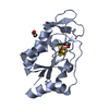 6z92SC  6z93C  6z94C  6zw9C  6z95 S: Starting model for refinement C: citing same article ( |
|---|---|
| Similar structure data |


-Links
 PDBj
PDBj- Assembly
| Deposited unit | 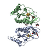
| ||||||||
|---|---|---|---|---|---|---|---|---|---|
| 1 | 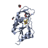
| ||||||||
| 2 | 
| ||||||||
| Unit cell |
|
-Components
-Protein , 1 types, 2 molecules AB
| #1: Protein | Mass: 17580.975 Da / Num. of mol.: 2 Source method: isolated from a genetically manipulated source Source: (gene. exp.) uncultured bacterium (environmental samples)Production host: |
|---|
-Non-polymers , 5 types, 342 molecules 
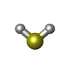


| #2: Chemical |  Mass: 351.640 Da / Num. of mol.: 2 / Source method: obtained synthetically / Formula: Fe4S4 / Feature type: SUBJECT OF INVESTIGATION Mass: 351.640 Da / Num. of mol.: 2 / Source method: obtained synthetically / Formula: Fe4S4 / Feature type: SUBJECT OF INVESTIGATION#3: Chemical |  Mass: 34.081 Da / Num. of mol.: 2 / Source method: obtained synthetically / Formula: H2S Mass: 34.081 Da / Num. of mol.: 2 / Source method: obtained synthetically / Formula: H2S#4: Chemical |  Mass: 62.068 Da / Num. of mol.: 3 / Source method: obtained synthetically / Formula: C2H6O2 Mass: 62.068 Da / Num. of mol.: 3 / Source method: obtained synthetically / Formula: C2H6O2#5: Chemical |  Mass: 106.120 Da / Num. of mol.: 2 / Source method: obtained synthetically / Formula: C4H10O3 Mass: 106.120 Da / Num. of mol.: 2 / Source method: obtained synthetically / Formula: C4H10O3#6: Water | ChemComp-HOH / | Mass: 18.015 Da / Num. of mol.: 333 / Source method: isolated from a natural source / Formula: H2O |
|---|
-Details
| Has ligand of interest | Y |
|---|
-Experimental details
-Experiment
| Experiment | Method: X-RAY DIFFRACTION / Number of used crystals: 1 |
|---|
- Sample preparation
| Crystal | Density Matthews: 3.24 Å3/Da / Density % sol: 62.07 % |
|---|---|
| Crystal grow | Temperature: 293 K / Method: vapor diffusion, hanging drop / pH: 5.5 Details: Anaeroby, 10 mM MgCl2, 0.1 M MES, pH 5.5, 7-9% PEG 6000 |
-Data collection
| Diffraction | Mean temperature: 100 K / Serial crystal experiment: N |
|---|---|
| Diffraction source | Source: SYNCHROTRON / Site: SOLEIL / Beamline: PROXIMA 2 / Wavelength: 0.98011 Å |
| Detector | Type: DECTRIS EIGER X 9M / Detector: PIXEL / Date: Feb 5, 2020 |
| Radiation | Protocol: MAD / Monochromatic (M) / Laue (L): M / Scattering type: x-ray |
| Radiation wavelength | Wavelength: 0.98011 Å / Relative weight: 1 |
| Reflection | Resolution: 1.33→39.69 Å / Num. obs: 69773 / % possible obs: 92.2 % / Redundancy: 6.8 % / CC1/2: 0.998 / Rmerge(I) obs: 0.077 / Rrim(I) all: 0.083 / Net I/σ(I): 12.2 |
| Reflection shell | Resolution: 1.33→1.5 Å / Redundancy: 6 % / Rmerge(I) obs: 1.151 / Mean I/σ(I) obs: 1.5 / Num. unique obs: 3489 / CC1/2: 0.548 / Rrim(I) all: 1.26 |
- Processing
| Software |
| ||||||||||||||||||||||||||||||||||||||||||||||||||||||||||||||||||
|---|---|---|---|---|---|---|---|---|---|---|---|---|---|---|---|---|---|---|---|---|---|---|---|---|---|---|---|---|---|---|---|---|---|---|---|---|---|---|---|---|---|---|---|---|---|---|---|---|---|---|---|---|---|---|---|---|---|---|---|---|---|---|---|---|---|---|---|
| Refinement | Method to determine structure: MOLECULAR REPLACEMENT Starting model: 6Z92 Resolution: 1.327→39.69 Å / Cor.coef. Fo:Fc: 0.967 / Cor.coef. Fo:Fc free: 0.958 / SU R Cruickshank DPI: 0.053 / Cross valid method: THROUGHOUT / SU R Blow DPI: 0.056 / SU Rfree Blow DPI: 0.057 / SU Rfree Cruickshank DPI: 0.055
| ||||||||||||||||||||||||||||||||||||||||||||||||||||||||||||||||||
| Displacement parameters | Biso mean: 25.37 Å2
| ||||||||||||||||||||||||||||||||||||||||||||||||||||||||||||||||||
| Refine analyze | Luzzati coordinate error obs: 0.17 Å | ||||||||||||||||||||||||||||||||||||||||||||||||||||||||||||||||||
| Refinement step | Cycle: LAST / Resolution: 1.327→39.69 Å
| ||||||||||||||||||||||||||||||||||||||||||||||||||||||||||||||||||
| Refine LS restraints |
| ||||||||||||||||||||||||||||||||||||||||||||||||||||||||||||||||||
| LS refinement shell | Resolution: 1.33→1.44 Å
|
