| 登録情報 | データベース: PDB / ID: 6z7u
|
|---|
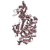
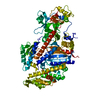
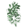
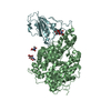
| タイトル | Myosin-II motor domain complexed with blebbistatin in a new ADP-release conformation |
|---|
 要素 要素 | Myosin-2 heavy chain |
|---|
 キーワード キーワード | MOTOR PROTEIN / myosin / motorprotein / blebbistatin / ADP-release / inhibitor / complex / hydrolase |
|---|
| 機能・相同性 |  機能・相同性情報 機能・相同性情報
uropod retraction / cytoplasmic actin-based contraction involved in forward cell motility / phagocytic cup base / pathogen-containing vacuole / response to differentiation-inducing factor 1 / equatorial cell cortex / contractile actin filament bundle assembly / pseudopodium retraction / cell trailing edge / contractile vacuole organization ...uropod retraction / cytoplasmic actin-based contraction involved in forward cell motility / phagocytic cup base / pathogen-containing vacuole / response to differentiation-inducing factor 1 / equatorial cell cortex / contractile actin filament bundle assembly / pseudopodium retraction / cell trailing edge / contractile vacuole organization / myosin filament assembly / aggregation involved in sorocarp development / culmination involved in sorocarp development / adenyl nucleotide binding / RHO GTPases activate PAKs / calcium-dependent ATPase activity / actomyosin contractile ring / hypotonic response / uropod / apical cortex / negative regulation of actin filament polymerization / actin-myosin filament sliding / detection of mechanical stimulus / substrate-dependent cell migration, cell extension / bleb assembly / actomyosin / filopodium assembly / early phagosome / myosin filament / cortical actin cytoskeleton organization / myosin II complex / cortical actin cytoskeleton / microfilament motor activity / cleavage furrow / pseudopodium / mitotic cytokinesis / cytoskeletal motor activity / response to cAMP / response to mechanical stimulus / 14-3-3 protein binding / response to hydrogen peroxide / cell motility / chemotaxis / actin filament binding / intracellular protein localization / regulation of cell shape / extracellular matrix / cytoplasmic vesicle / cell cortex / cytoskeleton / calmodulin binding / ATP binding / identical protein binding / cytoplasm / cytosol類似検索 - 分子機能 Methane Monooxygenase Hydroxylase; Chain G, domain 1 - #60 / Methane Monooxygenase Hydroxylase; Chain G, domain 1 - #530 / Myosin tail / Myosin tail / Myosin N-terminal SH3-like domain / Myosin S1 fragment, N-terminal / Myosin, N-terminal, SH3-like / Myosin N-terminal SH3-like domain profile. / Myosin motor domain profile. / Myosin head, motor domain ...Methane Monooxygenase Hydroxylase; Chain G, domain 1 - #60 / Methane Monooxygenase Hydroxylase; Chain G, domain 1 - #530 / Myosin tail / Myosin tail / Myosin N-terminal SH3-like domain / Myosin S1 fragment, N-terminal / Myosin, N-terminal, SH3-like / Myosin N-terminal SH3-like domain profile. / Myosin motor domain profile. / Myosin head, motor domain / Myosin head (motor domain) / Myosin. Large ATPases. / IQ motif profile. / Kinesin motor domain superfamily / Methane Monooxygenase Hydroxylase; Chain G, domain 1 / Up-down Bundle / P-loop containing nucleoside triphosphate hydrolase / Mainly Alpha類似検索 - ドメイン・相同性 ADENOSINE-5'-DIPHOSPHATE / Chem-BIT / Myosin-2 heavy chain類似検索 - 構成要素 |
|---|
| 生物種 |   Dictyostelium discoideum (キイロタマホコリカビ) Dictyostelium discoideum (キイロタマホコリカビ) |
|---|
| 手法 |  X線回折 / シンクロトロン / 分子置換 / 解像度: 2.58 Å X線回折 / シンクロトロン / 分子置換 / 解像度: 2.58 Å |
|---|
 データ登録者 データ登録者 | Ewert, W. / Preller, M. |
|---|
| 資金援助 |  ドイツ, 1件 ドイツ, 1件 | 組織 | 認可番号 | 国 |
|---|
| German Research Foundation (DFG) | PR 1478/2-1 | ドイツ |
|
|---|
 引用 引用 | ジャーナル: Int J Mol Sci / 年: 2020
タイトル: Structural and Computational Insights into a Blebbistatin-Bound Myosin•ADP Complex with Characteristics of an ADP-Release Conformation along the Two-Step Myosin Power Stoke.
著者: Ewert, W. / Franz, P. / Tsiavaliaris, G. / Preller, M. |
|---|
| 履歴 | | 登録 | 2020年6月1日 | 登録サイト: PDBE / 処理サイト: PDBE |
|---|
| 改定 1.0 | 2020年10月21日 | Provider: repository / タイプ: Initial release |
|---|
| 改定 1.1 | 2020年10月28日 | Group: Database references / カテゴリ: citation / citation_author
Item: _citation.journal_volume / _citation.pdbx_database_id_PubMed ..._citation.journal_volume / _citation.pdbx_database_id_PubMed / _citation.title / _citation_author.identifier_ORCID |
|---|
| 改定 1.2 | 2024年1月24日 | Group: Data collection / Database references / Refinement description
カテゴリ: chem_comp_atom / chem_comp_bond ...chem_comp_atom / chem_comp_bond / database_2 / pdbx_initial_refinement_model
Item: _database_2.pdbx_DOI / _database_2.pdbx_database_accession |
|---|
|
|---|
 ムービー
ムービー コントローラー


 データを開く
 基本情報
基本情報 構造の表示
構造の表示 ダウンロードとリンク
 その他のダウンロード
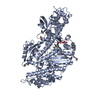


 PDBj
PDBj


 集合体
集合体

 分子量: 62.068 Da / 分子数: 9 / 由来タイプ: 合成 / 式: C2H6O2
 分子量: 427.201 Da / 分子数: 1 / 由来タイプ: 合成 / 式: C10H15N5O10P2 / タイプ: SUBJECT OF INVESTIGATION / コメント: ADP, エネルギー貯蔵分子*YM
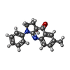
 分子量: 292.332 Da / 分子数: 1 / 由来タイプ: 合成 / 式: C18H16N2O2 / タイプ: SUBJECT OF INVESTIGATION
 分子量: 18.015 Da / 分子数: 59 / 由来タイプ: 天然 / 式: H2O
 試料調製
試料調製 / ビームライン: PROXIMA 2 / 波長: 0.9801 Å
 解析
解析