 Movie
Movie Controller

[English] 日本語
 Yorodumi
- PDB-4cs9: Crystal structure of the asymmetric human metapneumovirus M2-1 te... -
+ Open data

- Basic information
| Entry | Database: PDB / ID: 4cs9 | ||||||
|---|---|---|---|---|---|---|---|
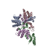
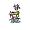
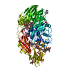
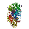
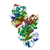
| Title | Crystal structure of the asymmetric human metapneumovirus M2-1 tetramer bound to adenosine monophosphate | ||||||
 Components Components | (M2-1) x 2 | ||||||
 Keywords Keywords | VIRAL PROTEIN / ANTITERMINATOR / TRANSCRIPTION ELONGATION / RNA-BINDING / MODULAR PROTEIN / ASYMMETRIC TETRAMER | ||||||
| Function / homology |  Function and homology information Function and homology informationregulation of viral transcription / virion component / host cell cytoplasm / nucleotide binding / host cell nucleus / structural molecule activity / RNA binding / zinc ion binding Similarity search - Function | ||||||
| Biological species |  HUMAN METAPNEUMOVIRUS HUMAN METAPNEUMOVIRUS | ||||||
| Method |  X-RAY DIFFRACTION / SYNCHROTRON / MOLECULAR REPLACEMENT / Resolution: 2.01 Å X-RAY DIFFRACTION / SYNCHROTRON / MOLECULAR REPLACEMENT / Resolution: 2.01 Å | ||||||
 Authors Authors | Leyrat, C. / Renner, M. / Harlos, K. / Grimes, J.M. | ||||||
 Citation Citation | Journal: Elife / Year: 2014 Title: Drastic Changes in Conformational Dynamics of the Antiterminator M2-1 Regulate Transcription Efficiency in Pneumovirinae. Authors: Leyrat, C. / Renner, M. / Harlos, K. / Huiskonen, J.T. / Grimes, J.M. | ||||||
| History |
|
- Structure visualization
| Structure viewer | Molecule: MolmilJmol/JSmol |
|---|
- Downloads & links
-Download
| PDBx/mmCIF format | 4cs9.cif.gz | 282.4 KB | Display | PDBx/mmCIF format |
|---|---|---|---|---|
| PDB format | pdb4cs9.ent.gz | 230.4 KB | Display | PDB format |
| PDBx/mmJSON format | 4cs9.json.gz | Tree view | PDBx/mmJSON format | |
| Others |  Other downloads Other downloads |
-Validation report
| Arichive directory | https://data.pdbj.org/pub/pdb/validation_reports/cs/4cs9ftp://data.pdbj.org/pub/pdb/validation_reports/cs/4cs9 | HTTPS FTP |
|---|
-Related structure data
| Related structure data |  4cs7C  4cs8SC  4csaC C: citing same article ( S: Starting model for refinement |
|---|---|
| Similar structure data |


-Links
 PDBj
PDBj


- Assembly
| Deposited unit | 
| ||||||||
|---|---|---|---|---|---|---|---|---|---|
| 1 |
| ||||||||
| Unit cell |
|
-Components
| #1: Protein | Mass: 21434.266 Da / Num. of mol.: 3 Source method: isolated from a genetically manipulated source Source: (gene. exp.) HUMAN METAPNEUMOVIRUS / Strain: NL1-00 (A1) / Plasmid: POPINF / Production host:  #2: Protein | | Mass: 21435.250 Da / Num. of mol.: 1 Source method: isolated from a genetically manipulated source Source: (gene. exp.) HUMAN METAPNEUMOVIRUS / Strain: NL1-00 (A1) / Plasmid: POPINF / Production host: #3: Chemical | ChemComp-ZN /   Mass: 65.409 Da / Num. of mol.: 4 / Source method: obtained synthetically / Formula: Zn Mass: 65.409 Da / Num. of mol.: 4 / Source method: obtained synthetically / Formula: Zn#4: Chemical | ChemComp-AMP /   Mass: 347.221 Da / Num. of mol.: 4 / Source method: obtained synthetically / Formula: C10H14N5O7P / Comment: AMP*YM Mass: 347.221 Da / Num. of mol.: 4 / Source method: obtained synthetically / Formula: C10H14N5O7P / Comment: AMP*YM#5: Water | ChemComp-HOH / |  Mass: 18.015 Da / Num. of mol.: 438 / Source method: isolated from a natural source / Formula: H2O Mass: 18.015 Da / Num. of mol.: 438 / Source method: isolated from a natural source / Formula: H2O |
|---|
-Experimental details
-Experiment
| Experiment | Method: X-RAY DIFFRACTION / Number of used crystals: 2 |
|---|
- Sample preparation
| Crystal | Density Matthews: 2.33 Å3/Da / Density % sol: 48.3 % / Description: NONE |
|---|---|
| Crystal grow | pH: 6.5 Details: 28 % W/V POLYETHYLENE GLYCOL MONOMETHYL ETHER 2000, 0.100 M BIS-TRIS PH 6.5 |
-Data collection
| Diffraction | Mean temperature: 100 K |
|---|---|
| Diffraction source | Source: SYNCHROTRON / Site: Diamond  / Beamline: I24 / Wavelength: 0.96862 / Beamline: I24 / Wavelength: 0.96862 |
| Detector | Type: DECTRIS PILATUS 6M / Detector: PIXEL / Date: Jul 12, 2013 / Details: MIRRORS |
| Radiation | Monochromator: DOUBLE CRYSTAL / Protocol: SINGLE WAVELENGTH / Monochromatic (M) / Laue (L): M / Scattering type: x-ray |
| Radiation wavelength | Wavelength: 0.96862 Å / Relative weight: 1 |
| Reflection | Resolution: 2.01→49.8 Å / Num. obs: 51659 / % possible obs: 98.8 % / Observed criterion σ(I): 1.8 / Redundancy: 12.8 % / Biso Wilson estimate: 42.15 Å2 / Rmerge(I) obs: 0.07 / Net I/σ(I): 22.5 |
| Reflection shell | Resolution: 2.01→2.06 Å / Redundancy: 5.1 % / Rmerge(I) obs: 0.84 / Mean I/σ(I) obs: 1.8 / % possible all: 85.1 |
- Processing
| Software |
| |||||||||||||||||||||||||||||||||||||||||||||||||||||||||||||||||||||||||||||||||||||||||||||||||||||||||||||||||||||||||||||
|---|---|---|---|---|---|---|---|---|---|---|---|---|---|---|---|---|---|---|---|---|---|---|---|---|---|---|---|---|---|---|---|---|---|---|---|---|---|---|---|---|---|---|---|---|---|---|---|---|---|---|---|---|---|---|---|---|---|---|---|---|---|---|---|---|---|---|---|---|---|---|---|---|---|---|---|---|---|---|---|---|---|---|---|---|---|---|---|---|---|---|---|---|---|---|---|---|---|---|---|---|---|---|---|---|---|---|---|---|---|---|---|---|---|---|---|---|---|---|---|---|---|---|---|---|---|---|
| Refinement | Method to determine structure: MOLECULAR REPLACEMENT Starting model: PDB ENTRY 4CS8 Resolution: 2.01→49.8 Å / Cor.coef. Fo:Fc: 0.9538 / Cor.coef. Fo:Fc free: 0.9425 / SU R Cruickshank DPI: 0.169 / Cross valid method: THROUGHOUT / σ(F): 0 / SU R Blow DPI: 0.172 / SU Rfree Blow DPI: 0.14 / SU Rfree Cruickshank DPI: 0.14 Details: THE RIBOSE AND PHOSPHATE OF AMP4 WERE MODELED STEREOCHEMICALLY
| |||||||||||||||||||||||||||||||||||||||||||||||||||||||||||||||||||||||||||||||||||||||||||||||||||||||||||||||||||||||||||||
| Displacement parameters | Biso mean: 53.74 Å2
| |||||||||||||||||||||||||||||||||||||||||||||||||||||||||||||||||||||||||||||||||||||||||||||||||||||||||||||||||||||||||||||
| Refine analyze | Luzzati coordinate error obs: 0.28 Å | |||||||||||||||||||||||||||||||||||||||||||||||||||||||||||||||||||||||||||||||||||||||||||||||||||||||||||||||||||||||||||||
| Refinement step | Cycle: LAST / Resolution: 2.01→49.8 Å
| |||||||||||||||||||||||||||||||||||||||||||||||||||||||||||||||||||||||||||||||||||||||||||||||||||||||||||||||||||||||||||||
| Refine LS restraints |
| |||||||||||||||||||||||||||||||||||||||||||||||||||||||||||||||||||||||||||||||||||||||||||||||||||||||||||||||||||||||||||||
| LS refinement shell | Resolution: 2.01→2.06 Å / Total num. of bins used: 20
| |||||||||||||||||||||||||||||||||||||||||||||||||||||||||||||||||||||||||||||||||||||||||||||||||||||||||||||||||||||||||||||
| Refinement TLS params. | Method: refined / Refine-ID: X-RAY DIFFRACTION
| |||||||||||||||||||||||||||||||||||||||||||||||||||||||||||||||||||||||||||||||||||||||||||||||||||||||||||||||||||||||||||||
| Refinement TLS group |
|
