 Movie
Movie Controller

[English] 日本語
 Yorodumi
- PDB-1xc6: Native Structure Of Beta-Galactosidase from Penicillium sp. in co... -
+ Open data

- Basic information
| Entry | Database: PDB / ID: 1xc6 | |||||||||
|---|---|---|---|---|---|---|---|---|---|---|
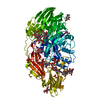
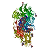
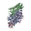
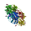
| Title | Native Structure Of Beta-Galactosidase from Penicillium sp. in complex with Galactose | |||||||||
 Components Components | Beta-Galactosidase | |||||||||
 Keywords Keywords | HYDROLASE / Tim Barrel Domain / Glycoside Hydrolase / Family GH35 / Glycoprotein / Penicillium / Quick Cryo Soaking | |||||||||
| Function / homology |  Function and homology information Function and homology informationgalactose binding / lactose catabolic process / beta-galactosidase / beta-galactosidase activity / polysaccharide catabolic process / : / extracellular region Similarity search - Function | |||||||||
| Biological species |  Penicillium sp. (fungus) Penicillium sp. (fungus) | |||||||||
| Method |  X-RAY DIFFRACTION / SYNCHROTRON / MIR / Resolution: 2.1 Å X-RAY DIFFRACTION / SYNCHROTRON / MIR / Resolution: 2.1 Å | |||||||||
 Authors Authors | Rojas, A.L. / Nagem, R.A.P. / Neustroev, K.N. / Arand, M. / Adamska, M. / Eneyskaya, E.V. / Kulminskaya, A.A. / Garratt, R.C. / Golubev, A.M. / Polikarpov, I. | |||||||||
 Citation Citation | Journal: J.Mol.Biol. / Year: 2004 Title: Crystal Structures of beta-Galactosidase from Penicillium sp. and its Complex with Galactose Authors: Rojas, A.L. / Nagem, R.A.P. / Neustroev, K.N. / Arand, M. / Adamska, M. / Eneyskaya, E.V. / Kulminskaya, A.A. / Garratt, R.C. / Golubev, A.M. / Polikarpov, I. #1: Journal: Acta Crystallogr.,Sect.D / Year: 2000Title: purification, crystallization and preliminary diffraction study of beta-galactosidase from Penicillium sp. Authors: Neustroev, K.N. / De Sousa, E.A. / Golubev, A.M. / Brandao Neto, J.R. / Eneyskaya, E.V. / Kulminskaya, A.A. / Polikarpov, I. | |||||||||
| History |
|
- Structure visualization
| Structure viewer | Molecule: MolmilJmol/JSmol |
|---|
- Downloads & links
-Download
| PDBx/mmCIF format | 1xc6.cif.gz | 239 KB | Display | PDBx/mmCIF format |
|---|---|---|---|---|
| PDB format | pdb1xc6.ent.gz | 184.7 KB | Display | PDB format |
| PDBx/mmJSON format | 1xc6.json.gz | Tree view | PDBx/mmJSON format | |
| Others |  Other downloads Other downloads |
-Validation report
| Arichive directory | https://data.pdbj.org/pub/pdb/validation_reports/xc/1xc6ftp://data.pdbj.org/pub/pdb/validation_reports/xc/1xc6 | HTTPS FTP |
|---|
-Related structure data


-Links
 PDBj
PDBj


- Assembly
| Deposited unit | 
| ||||||||
|---|---|---|---|---|---|---|---|---|---|
| 1 |
| ||||||||
| Unit cell |
| ||||||||
| Details | The biological assembly is a monomer generated from the monomer in the asymetric unit. |
-Components
-Protein , 1 types, 1 molecules A
| #1: Protein | Mass: 105683.844 Da / Num. of mol.: 1 / Fragment: mature peptide (residues 41-1011) / Source method: isolated from a natural source / Source: (natural) Penicillium sp. (fungus)References: GenBank: 44844271, UniProt: Q700S9*PLUS, beta-galactosidase |
|---|
-Sugars , 7 types, 8 molecules 

| #2: Polysaccharide | beta-D-mannopyranose-(1-4)-2-acetamido-2-deoxy-beta-D-glucopyranose-(1-4)-2-acetamido-2-deoxy-beta- ...beta-D-mannopyranose-(1-4)-2-acetamido-2-deoxy-beta-D-glucopyranose-(1-4)-2-acetamido-2-deoxy-beta-D-glucopyranose Source method: isolated from a genetically manipulated source | ||
|---|---|---|---|
| #3: Polysaccharide | alpha-D-mannopyranose-(1-2)-alpha-D-mannopyranose-(1-3)-[alpha-D-mannopyranose-(1-2)-alpha-D- ...alpha-D-mannopyranose-(1-2)-alpha-D-mannopyranose-(1-3)-[alpha-D-mannopyranose-(1-2)-alpha-D-mannopyranose-(1-6)]alpha-D-mannopyranose-(1-6)-[beta-D-mannopyranose-(1-3)]beta-D-mannopyranose-(1-4)-2-acetamido-2-deoxy-beta-D-glucopyranose-(1-4)-2-acetamido-2-deoxy-beta-D-glucopyranose Source method: isolated from a genetically manipulated source | ||
| #4: Polysaccharide | 2-acetamido-2-deoxy-beta-D-glucopyranose-(1-4)-2-acetamido-2-deoxy-beta-D-glucopyranose Source method: isolated from a genetically manipulated source | ||
| #5: Polysaccharide | alpha-D-mannopyranose-(1-3)-alpha-D-mannopyranose-(1-6)-beta-D-mannopyranose-(1-4)-2-acetamido-2- ...alpha-D-mannopyranose-(1-3)-alpha-D-mannopyranose-(1-6)-beta-D-mannopyranose-(1-4)-2-acetamido-2-deoxy-beta-D-glucopyranose-(1-4)-2-acetamido-2-deoxy-beta-D-glucopyranose Source method: isolated from a genetically manipulated source | ||
| #6: Polysaccharide | alpha-D-mannopyranose-(1-3)-[alpha-D-mannopyranose-(1-6)]alpha-D-mannopyranose-(1-6)-[alpha-D- ...alpha-D-mannopyranose-(1-3)-[alpha-D-mannopyranose-(1-6)]alpha-D-mannopyranose-(1-6)-[alpha-D-mannopyranose-(1-3)]beta-D-mannopyranose-(1-4)-2-acetamido-2-deoxy-beta-D-glucopyranose-(1-4)-2-acetamido-2-deoxy-beta-D-glucopyranose Source method: isolated from a genetically manipulated source | ||
| #7: Sugar |  Type: D-saccharide, beta linking / Mass: 221.208 Da / Num. of mol.: 2 Type: D-saccharide, beta linking / Mass: 221.208 Da / Num. of mol.: 2Source method: isolated from a genetically manipulated source Formula: C8H15NO6 #8: Sugar | ChemComp-GAL / |  Type: D-saccharide, beta linking / Mass: 180.156 Da / Num. of mol.: 1 Type: D-saccharide, beta linking / Mass: 180.156 Da / Num. of mol.: 1Source method: isolated from a genetically manipulated source Formula: C6H12O6 |
-Non-polymers , 5 types, 1017 molecules 


| #9: Chemical | ChemComp-NA /  Mass: 22.990 Da / Num. of mol.: 1 / Source method: obtained synthetically / Formula: Na Mass: 22.990 Da / Num. of mol.: 1 / Source method: obtained synthetically / Formula: Na | ||||
|---|---|---|---|---|---|
| #10: Chemical | ChemComp-PO4 /  Mass: 94.971 Da / Num. of mol.: 1 / Source method: obtained synthetically / Formula: PO4 Mass: 94.971 Da / Num. of mol.: 1 / Source method: obtained synthetically / Formula: PO4 | ||||
| #11: Chemical | ChemComp-IOD /  Mass: 126.904 Da / Num. of mol.: 52 / Source method: obtained synthetically / Formula: I Mass: 126.904 Da / Num. of mol.: 52 / Source method: obtained synthetically / Formula: I#12: Chemical | ChemComp-EDO /  Mass: 62.068 Da / Num. of mol.: 11 / Source method: obtained synthetically / Formula: C2H6O2 Mass: 62.068 Da / Num. of mol.: 11 / Source method: obtained synthetically / Formula: C2H6O2#13: Water | ChemComp-HOH / | Mass: 18.015 Da / Num. of mol.: 952 / Source method: isolated from a natural source / Formula: H2O |
-Details
| Has protein modification | Y |
|---|
-Experimental details
-Experiment
| Experiment | Method: X-RAY DIFFRACTION / Number of used crystals: 1 |
|---|
- Sample preparation
| Crystal | Density Matthews: 4.5 Å3/Da / Density % sol: 72.5 % |
|---|---|
| Crystal grow | Temperature: 291 K / Method: vapor diffusion, hanging drop / pH: 4 Details: 15% PEG 8000, 50 MM Sodium Phosphate (Iodide ions were incorporated into the crystal by Quick Cryo Soaking technique at room temperature), pH 4.0, Vapor Diffusion, Hanging Drop, temperature 291K |
-Data collection
| Diffraction | Mean temperature: 100 K |
|---|---|
| Diffraction source | Source: SYNCHROTRON / Site: LNLS  / Beamline: D03B-MX1 / Wavelength: 1.54 Å / Beamline: D03B-MX1 / Wavelength: 1.54 Å |
| Detector | Type: MARRESEARCH / Detector: IMAGE PLATE / Date: Jan 1, 2000 |
| Radiation | Protocol: SINGLE WAVELENGTH / Monochromatic (M) / Laue (L): M / Scattering type: x-ray |
| Radiation wavelength | Wavelength: 1.54 Å / Relative weight: 1 |
| Reflection | Resolution: 2.1→23.4 Å / Num. all: 224036 / Num. obs: 224036 / % possible obs: 99.9 % / Observed criterion σ(F): 0 / Observed criterion σ(I): 3.1 / Redundancy: 4.2 % / Rmerge(I) obs: 0.107 / Net I/σ(I): 12.5 |
| Reflection shell | Resolution: 2.1→2.15 Å / Redundancy: 4.2 % / Rmerge(I) obs: 0.481 / Mean I/σ(I) obs: 3.1 / % possible all: 100 |
- Processing
| Software |
| |||||||||||||||||||||||||
|---|---|---|---|---|---|---|---|---|---|---|---|---|---|---|---|---|---|---|---|---|---|---|---|---|---|---|
| Refinement | Method to determine structure: MIR / Resolution: 2.1→23.4 Å / Isotropic thermal model: ISOTROPIC / σ(F): 0 / σ(I): 3.1 / Stereochemistry target values: Engh & Huber
| |||||||||||||||||||||||||
| Displacement parameters | Biso mean: 22.63 Å2 | |||||||||||||||||||||||||
| Refine analyze |
| |||||||||||||||||||||||||
| Refinement step | Cycle: LAST / Resolution: 2.1→23.4 Å
| |||||||||||||||||||||||||
| Refine LS restraints |
|
