 Movie
Movie Controller


+ Open data

- Basic information
| Entry | Database: PDB / ID: 6z15 | ||||||
|---|---|---|---|---|---|---|---|
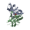
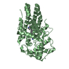
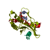
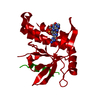
| Title | Human wtSTING in complex with 3',3'-c-di-AMP | ||||||
 Components Components | Stimulator of interferon protein | ||||||
 Keywords Keywords | PROTEIN BINDING / CDN / Innate immune system / STING / activator | ||||||
| Function / homology |  Function and homology information Function and homology informationautophagosome membrane / positive regulation of type I interferon production / endoplasmic reticulum-Golgi intermediate compartment membrane / activation of innate immune response / mitochondrial outer membrane / nucleotide binding / endoplasmic reticulum membrane / perinuclear region of cytoplasm Similarity search - Function | ||||||
| Biological species |  Homo sapiens (human) Homo sapiens (human) | ||||||
| Method |  X-RAY DIFFRACTION / SYNCHROTRON / MOLECULAR REPLACEMENT / molecular replacement / Resolution: 2.5 Å X-RAY DIFFRACTION / SYNCHROTRON / MOLECULAR REPLACEMENT / molecular replacement / Resolution: 2.5 Å | ||||||
 Authors Authors | Boura, E. / Smola, M. | ||||||
 Citation Citation | Journal: Angew.Chem.Int.Ed.Engl. / Year: 2021 Title: Ligand Strain and Its Conformational Complexity Is a Major Factor in the Binding of Cyclic Dinucleotides to STING Protein. Authors: Smola, M. / Gutten, O. / Dejmek, M. / Kozisek, M. / Evangelidis, T. / Tehrani, Z.A. / Novotna, B. / Nencka, R. / Birkus, G. / Rulisek, L. / Boura, E. | ||||||
| History |
|
- Structure visualization
| Structure viewer | Molecule: MolmilJmol/JSmol |
|---|
- Downloads & links
-Download
| PDBx/mmCIF format | 6z15.cif.gz | 82.2 KB | Display | PDBx/mmCIF format |
|---|---|---|---|---|
| PDB format | pdb6z15.ent.gz | 60.7 KB | Display | PDB format |
| PDBx/mmJSON format | 6z15.json.gz | Tree view | PDBx/mmJSON format | |
| Others |  Other downloads Other downloads |
-Validation report
| Summary document | 6z15_validation.pdf.gz | 835.7 KB | Display | wwPDB validaton report |
|---|---|---|---|---|
| Full document | 6z15_full_validation.pdf.gz | 836.2 KB | Display | |
| Data in XML | 6z15_validation.xml.gz | 9.1 KB | Display | |
| Data in CIF | 6z15_validation.cif.gz | 11.6 KB | Display | |
| Arichive directory | https://data.pdbj.org/pub/pdb/validation_reports/z1/6z15ftp://data.pdbj.org/pub/pdb/validation_reports/z1/6z15 | HTTPS FTP |
-Related structure data
| Related structure data |  6y99C 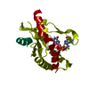 6ydbC  6ydzC 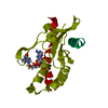 6yeaC  6z0zC  4ksyS S: Starting model for refinement C: citing same article ( |
|---|---|
| Similar structure data |


-Links
 PDBj
PDBj- Assembly
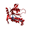
| Deposited unit | 
| ||||||||
|---|---|---|---|---|---|---|---|---|---|
| 1 | 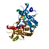
| ||||||||
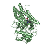
| Unit cell |
|
-Components
| #1: Protein | Mass: 23189.064 Da / Num. of mol.: 1 Source method: isolated from a genetically manipulated source Source: (gene. exp.) Homo sapiens (human) / Gene: STING, LOC340061, hCG_1782396 / Production host:  |
|---|---|
| #2: Chemical | ChemComp-2BA / (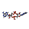  Mass: 658.412 Da / Num. of mol.: 1 / Source method: obtained synthetically / Formula: C20H24N10O12P2 / Feature type: SUBJECT OF INVESTIGATION Mass: 658.412 Da / Num. of mol.: 1 / Source method: obtained synthetically / Formula: C20H24N10O12P2 / Feature type: SUBJECT OF INVESTIGATION |
| #3: Water | ChemComp-HOH /  Mass: 18.015 Da / Num. of mol.: 30 / Source method: isolated from a natural source / Formula: H2O Mass: 18.015 Da / Num. of mol.: 30 / Source method: isolated from a natural source / Formula: H2O |
| Has ligand of interest | Y |
-Experimental details
-Experiment
| Experiment | Method: X-RAY DIFFRACTION / Number of used crystals: 1 |
|---|
- Sample preparation
| Crystal | Density Matthews: 2.35 Å3/Da / Density % sol: 47.75 % |
|---|---|
| Crystal grow | Temperature: 293 K / Method: vapor diffusion, sitting drop Details: 12.5% w/v PEG 1000, 12.5% w/v PEG 3350, 12.5% v/v MPD, 0.1 M bicine/Trizma base pH 8.5, 0.03 M diethyleneglycol, 0.03 M triethyleneglycol, 0.03 M tetraethyleneglycol, 0.03 M pentaethyleneglycol |
-Data collection
| Diffraction | Mean temperature: 100 K / Serial crystal experiment: N |
|---|---|
| Diffraction source | Source: SYNCHROTRON / Site: BESSY  / Beamline: 14.2 / Wavelength: 0.9184 Å / Beamline: 14.2 / Wavelength: 0.9184 Å |
| Detector | Type: DECTRIS PILATUS 2M / Detector: PIXEL / Date: Apr 6, 2018 |
| Radiation | Protocol: SINGLE WAVELENGTH / Monochromatic (M) / Laue (L): M / Scattering type: x-ray |
| Radiation wavelength | Wavelength: 0.9184 Å / Relative weight: 1 |
| Reflection | Resolution: 2.196→39.2 Å / Num. obs: 11816 / % possible obs: 99.82 % / Redundancy: 13.6 % / CC1/2: 0.998 / Net I/σ(I): 10.89 |
| Reflection shell | Resolution: 2.196→2.275 Å / Mean I/σ(I) obs: 0.56 / Num. unique obs: 1138 / CC1/2: 0.245 |
-Phasing
| Phasing | Method: molecular replacement |
|---|
- Processing
| Software |
| ||||||||||||||||||||||||
|---|---|---|---|---|---|---|---|---|---|---|---|---|---|---|---|---|---|---|---|---|---|---|---|---|---|
| Refinement | Method to determine structure: MOLECULAR REPLACEMENT Starting model: 4KSY Resolution: 2.5→39.1389 Å / SU ML: 0.29 / Cross valid method: THROUGHOUT / σ(F): 1.37 / Phase error: 27.58 / Stereochemistry target values: ML
| ||||||||||||||||||||||||
| Solvent computation | Shrinkage radii: 0.9 Å / VDW probe radii: 1.11 Å / Solvent model: FLAT BULK SOLVENT MODEL | ||||||||||||||||||||||||
| Displacement parameters | Biso max: 129.84 Å2 / Biso mean: 61.1017 Å2 / Biso min: 29.15 Å2 | ||||||||||||||||||||||||
| Refinement step | Cycle: final / Resolution: 2.5→39.1389 Å
| ||||||||||||||||||||||||
| LS refinement shell | Refine-ID: X-RAY DIFFRACTION / Rfactor Rfree error: 0 / % reflection obs: 100 %
|
