 Movie
Movie Controller


+ Open data

- Basic information
| Entry | Database: PDB / ID: 6cy7 | ||||||
|---|---|---|---|---|---|---|---|
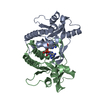
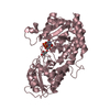
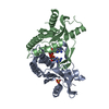
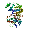
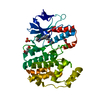
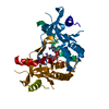
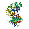
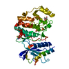
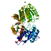
| Title | Human Stimulator of Interferon Genes | ||||||
 Components Components | Stimulator of interferon genes protein | ||||||
 Keywords Keywords | IMMUNE SYSTEM / human STING / complex / cyclic-di-AMP / TMEM173 / Ala230 allelle / 230A/232R | ||||||
| Function / homology |  Function and homology information Function and homology informationSTAT6-mediated induction of chemokines / protein localization to endoplasmic reticulum / 2',3'-cyclic GMP-AMP binding / cyclic-di-GMP binding / STING mediated induction of host immune responses / Dengue virus activates/modulates innate and adaptive immune responses / positive regulation of type I interferon-mediated signaling pathway / IRF3-mediated induction of type I IFN / proton channel activity / reticulophagy ...STAT6-mediated induction of chemokines / protein localization to endoplasmic reticulum / 2',3'-cyclic GMP-AMP binding / cyclic-di-GMP binding / STING mediated induction of host immune responses / Dengue virus activates/modulates innate and adaptive immune responses / positive regulation of type I interferon-mediated signaling pathway / IRF3-mediated induction of type I IFN / proton channel activity / reticulophagy / cGAS/STING signaling pathway / pattern recognition receptor signaling pathway / cellular response to exogenous dsRNA / cytoplasmic pattern recognition receptor signaling pathway / protein complex oligomerization / positive regulation of macroautophagy / autophagosome membrane / autophagosome assembly / positive regulation of defense response to virus by host / positive regulation of type I interferon production / endoplasmic reticulum-Golgi intermediate compartment membrane / activation of innate immune response / signaling adaptor activity / positive regulation of interferon-beta production / autophagosome / cytoplasmic vesicle membrane / secretory granule membrane / Regulation of innate immune responses to cytosolic DNA / antiviral innate immune response / protein serine/threonine kinase binding / SARS-CoV-1 activates/modulates innate immune responses / peroxisome / defense response to virus / RNA polymerase II-specific DNA-binding transcription factor binding / transcription coactivator activity / mitochondrial outer membrane / endosome / Golgi membrane / innate immune response / ubiquitin protein ligase binding / Neutrophil degranulation / protein kinase binding / endoplasmic reticulum membrane / perinuclear region of cytoplasm / SARS-CoV-2 activates/modulates innate and adaptive immune responses / protein homodimerization activity / positive regulation of transcription by RNA polymerase II / identical protein binding / plasma membrane / cytosol Similarity search - Function | ||||||
| Biological species |  Homo sapiens (human) Homo sapiens (human) | ||||||
| Method |  X-RAY DIFFRACTION / SYNCHROTRON / MOLECULAR REPLACEMENT / Resolution: 2.2 Å X-RAY DIFFRACTION / SYNCHROTRON / MOLECULAR REPLACEMENT / Resolution: 2.2 Å | ||||||
 Authors Authors | Fernandez, D. / Li, L. / Ergun, S.L. | ||||||
 Citation Citation | Journal: Cell / Year: 2019 Title: STING Polymer Structure Reveals Mechanisms for Activation, Hyperactivation, and Inhibition. Authors: Ergun, S.L. / Fernandez, D. / Weiss, T.M. / Li, L. #1: Journal: Biorxiv / Year: 2019Title: STING polymer structure reveals mechanisms for activation, hyperactivation, and inhibition Authors: Ergun, S.L. / Fernandez, D. / Weiss, T.M. / Li, L. | ||||||
| History |
|
- Structure visualization
| Structure viewer | Molecule: MolmilJmol/JSmol |
|---|
- Downloads & links
-Download
| PDBx/mmCIF format | 6cy7.cif.gz | 57.3 KB | Display | PDBx/mmCIF format |
|---|---|---|---|---|
| PDB format | pdb6cy7.ent.gz | 38 KB | Display | PDB format |
| PDBx/mmJSON format | 6cy7.json.gz | Tree view | PDBx/mmJSON format | |
| Others |  Other downloads Other downloads |
-Validation report
| Arichive directory | https://data.pdbj.org/pub/pdb/validation_reports/cy/6cy7ftp://data.pdbj.org/pub/pdb/validation_reports/cy/6cy7 | HTTPS FTP |
|---|
-Related structure data
| Related structure data | 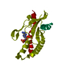 6cffC 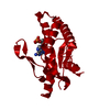 6dnkC  4lohS S: Starting model for refinement C: citing same article ( |
|---|---|
| Similar structure data |

-Links
 PDBj
PDBj


- Assembly
| Deposited unit | 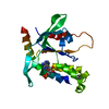
| ||||||||
|---|---|---|---|---|---|---|---|---|---|
| 1 | 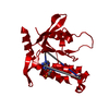
| ||||||||
| Unit cell |
| ||||||||
| Components on special symmetry positions |
|
-Components
| #1: Protein | Mass: 27137.619 Da / Num. of mol.: 1 / Fragment: UNP residues 139-379 Source method: isolated from a genetically manipulated source Source: (gene. exp.) Homo sapiens (human) / Gene: TMEM173, ERIS, MITA, STING / Production host:  |
|---|---|
| #2: Chemical | ChemComp-2BA / (  Mass: 658.412 Da / Num. of mol.: 1 / Source method: obtained synthetically / Formula: C20H24N10O12P2 / Feature type: SUBJECT OF INVESTIGATION Mass: 658.412 Da / Num. of mol.: 1 / Source method: obtained synthetically / Formula: C20H24N10O12P2 / Feature type: SUBJECT OF INVESTIGATION |
| #3: Chemical | ChemComp-IMD /   Mass: 69.085 Da / Num. of mol.: 1 / Source method: obtained synthetically / Formula: C3H5N2 Mass: 69.085 Da / Num. of mol.: 1 / Source method: obtained synthetically / Formula: C3H5N2 |
| #4: Chemical | ChemComp-GOL /   Mass: 92.094 Da / Num. of mol.: 1 / Source method: obtained synthetically / Formula: C3H8O3 Mass: 92.094 Da / Num. of mol.: 1 / Source method: obtained synthetically / Formula: C3H8O3 |
| #5: Water | ChemComp-HOH /  Mass: 18.015 Da / Num. of mol.: 51 / Source method: isolated from a natural source / Formula: H2O Mass: 18.015 Da / Num. of mol.: 51 / Source method: isolated from a natural source / Formula: H2O |
-Experimental details
-Experiment
| Experiment | Method: X-RAY DIFFRACTION / Number of used crystals: 1 |
|---|
- Sample preparation
| Crystal | Density Matthews: 2.03 Å3/Da / Density % sol: 39.32 % / Description: Rod |
|---|---|
| Crystal grow | Temperature: 285 K / Method: vapor diffusion, sitting drop / pH: 7 / Details: ammonium citrate tribasic, imidazole, PEG5000 MME |
-Data collection
| Diffraction | Mean temperature: 100 K |
|---|---|
| Diffraction source | Source: SYNCHROTRON / Site: SSRL  / Beamline: BL12-2 / Wavelength: 0.97946 Å / Beamline: BL12-2 / Wavelength: 0.97946 Å |
| Detector | Type: DECTRIS PILATUS 6M / Detector: PIXEL / Date: Mar 26, 2018 |
| Radiation | Monochromator: Liquid nitrogen-cooled double-crystal Si(111) Protocol: SINGLE WAVELENGTH / Monochromatic (M) / Laue (L): M / Scattering type: x-ray |
| Radiation wavelength | Wavelength: 0.97946 Å / Relative weight: 1 |
| Reflection | Resolution: 2.2→37 Å / Num. obs: 11912 / % possible obs: 99.9 % / Redundancy: 5.7 % / Biso Wilson estimate: 43 Å2 / CC1/2: 0.999 / Rmerge(I) obs: 0.062 / Rpim(I) all: 0.029 / Net I/σ(I): 14.2 |
| Reflection shell | Resolution: 2.2→2.32 Å / Redundancy: 5.5 % / Rmerge(I) obs: 0.657 / Mean I/σ(I) obs: 2.6 / Num. unique obs: 1696 / CC1/2: 0.759 / Rpim(I) all: 0.305 / Rrim(I) all: 0.726 / % possible all: 100 |
- Processing
| Software |
| ||||||||||||||||||||||||||||||||||||||||||||||||||||||||||||||||||||||||||||||||||||||||||||||||||||||||||||||||||||||||||||||||||||||||||||||||||||||||||||||||||||||||||||||||||||||
|---|---|---|---|---|---|---|---|---|---|---|---|---|---|---|---|---|---|---|---|---|---|---|---|---|---|---|---|---|---|---|---|---|---|---|---|---|---|---|---|---|---|---|---|---|---|---|---|---|---|---|---|---|---|---|---|---|---|---|---|---|---|---|---|---|---|---|---|---|---|---|---|---|---|---|---|---|---|---|---|---|---|---|---|---|---|---|---|---|---|---|---|---|---|---|---|---|---|---|---|---|---|---|---|---|---|---|---|---|---|---|---|---|---|---|---|---|---|---|---|---|---|---|---|---|---|---|---|---|---|---|---|---|---|---|---|---|---|---|---|---|---|---|---|---|---|---|---|---|---|---|---|---|---|---|---|---|---|---|---|---|---|---|---|---|---|---|---|---|---|---|---|---|---|---|---|---|---|---|---|---|---|---|---|
| Refinement | Method to determine structure: MOLECULAR REPLACEMENT Starting model: PDB entry 4LOH Resolution: 2.2→20.01 Å / Cor.coef. Fo:Fc: 0.96 / Cor.coef. Fo:Fc free: 0.939 / SU B: 7.045 / SU ML: 0.175 / Cross valid method: THROUGHOUT / ESU R: 0.245 / ESU R Free: 0.212 / Details: HYDROGENS HAVE BEEN ADDED IN THE RIDING POSITIONS
| ||||||||||||||||||||||||||||||||||||||||||||||||||||||||||||||||||||||||||||||||||||||||||||||||||||||||||||||||||||||||||||||||||||||||||||||||||||||||||||||||||||||||||||||||||||||
| Solvent computation | Ion probe radii: 0.8 Å / Shrinkage radii: 0.8 Å / VDW probe radii: 1.2 Å | ||||||||||||||||||||||||||||||||||||||||||||||||||||||||||||||||||||||||||||||||||||||||||||||||||||||||||||||||||||||||||||||||||||||||||||||||||||||||||||||||||||||||||||||||||||||
| Displacement parameters | Biso mean: 52.322 Å2
| ||||||||||||||||||||||||||||||||||||||||||||||||||||||||||||||||||||||||||||||||||||||||||||||||||||||||||||||||||||||||||||||||||||||||||||||||||||||||||||||||||||||||||||||||||||||
| Refinement step | Cycle: 1 / Resolution: 2.2→20.01 Å
| ||||||||||||||||||||||||||||||||||||||||||||||||||||||||||||||||||||||||||||||||||||||||||||||||||||||||||||||||||||||||||||||||||||||||||||||||||||||||||||||||||||||||||||||||||||||
| Refine LS restraints |
|