 Movie
Movie Controller

[English] 日本語
 Yorodumi
- PDB-6yxt: Crystal structure of the ADP-bound form of choline kinase from Pl... -
+ Open data

- Basic information
| Entry | Database: PDB / ID: 6yxt | ||||||
|---|---|---|---|---|---|---|---|
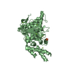
| Title | Crystal structure of the ADP-bound form of choline kinase from Plasmodium falciparum | ||||||
 Components Components | Choline kinase | ||||||
 Keywords Keywords | TRANSFERASE / choline kinase / ADP / complex | ||||||
| Function / homology |  Function and homology information Function and homology informationSynthesis of PE / choline kinase / ethanolamine kinase activity / choline kinase activity / Synthesis of PC / phosphatidylethanolamine biosynthetic process / phosphatidylcholine biosynthetic process / choline binding / nucleotide binding / metal ion binding ...Synthesis of PE / choline kinase / ethanolamine kinase activity / choline kinase activity / Synthesis of PC / phosphatidylethanolamine biosynthetic process / phosphatidylcholine biosynthetic process / choline binding / nucleotide binding / metal ion binding / cytoplasm / cytosol Similarity search - Function | ||||||
| Biological species |  | ||||||
| Method |  X-RAY DIFFRACTION / SYNCHROTRON / MOLECULAR REPLACEMENT / Resolution: 2.2 Å X-RAY DIFFRACTION / SYNCHROTRON / MOLECULAR REPLACEMENT / Resolution: 2.2 Å | ||||||
 Authors Authors | Torretta, A. / Parisini, E. | ||||||
 Citation Citation | Journal: Crystals / Year: 2020 Title: Crystal Structure of the Apo and the ADP-Bound Form of Choline Kinase from Plasmodium falciparum Authors: Torretta, A. / Lopez-Cara, L.C. / Parisini, E. | ||||||
| History |
|
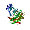
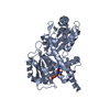
- Structure visualization
| Structure viewer | Molecule: MolmilJmol/JSmol |
|---|
- Downloads & links
-Download
| PDBx/mmCIF format | 6yxt.cif.gz | 95.9 KB | Display | PDBx/mmCIF format |
|---|---|---|---|---|
| PDB format | pdb6yxt.ent.gz | 69.6 KB | Display | PDB format |
| PDBx/mmJSON format | 6yxt.json.gz | Tree view | PDBx/mmJSON format | |
| Others |  Other downloads Other downloads |
-Validation report
| Arichive directory | https://data.pdbj.org/pub/pdb/validation_reports/yx/6yxtftp://data.pdbj.org/pub/pdb/validation_reports/yx/6yxt | HTTPS FTP |
|---|
-Related structure data
| Related structure data |  6yxsC 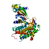 3fi8S C: citing same article ( S: Starting model for refinement |
|---|---|
| Similar structure data |


-Links
 PDBj
PDBj- Assembly
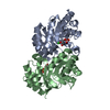
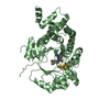
| Deposited unit | 
| ||||||||
|---|---|---|---|---|---|---|---|---|---|
| 1 |
| ||||||||
| Unit cell |
|
-Components
| #1: Protein | Mass: 45334.473 Da / Num. of mol.: 1 Source method: isolated from a genetically manipulated source Source: (gene. exp.) Gene: PF3D7_1401800 / Production host:  | ||||||||
|---|---|---|---|---|---|---|---|---|---|
| #2: Chemical |   Mass: 24.305 Da / Num. of mol.: 2 / Source method: obtained synthetically / Formula: Mg / Feature type: SUBJECT OF INVESTIGATION Mass: 24.305 Da / Num. of mol.: 2 / Source method: obtained synthetically / Formula: Mg / Feature type: SUBJECT OF INVESTIGATION#3: Chemical | ChemComp-ADP / |   Mass: 427.201 Da / Num. of mol.: 1 / Source method: obtained synthetically / Formula: C10H15N5O10P2 / Feature type: SUBJECT OF INVESTIGATION / Comment: ADP, energy-carrying molecule*YM Mass: 427.201 Da / Num. of mol.: 1 / Source method: obtained synthetically / Formula: C10H15N5O10P2 / Feature type: SUBJECT OF INVESTIGATION / Comment: ADP, energy-carrying molecule*YM#4: Chemical |   Mass: 62.068 Da / Num. of mol.: 3 / Source method: obtained synthetically / Formula: C2H6O2 Mass: 62.068 Da / Num. of mol.: 3 / Source method: obtained synthetically / Formula: C2H6O2#5: Water | ChemComp-HOH / |  Mass: 18.015 Da / Num. of mol.: 96 / Source method: isolated from a natural source / Formula: H2O Mass: 18.015 Da / Num. of mol.: 96 / Source method: isolated from a natural source / Formula: H2OHas ligand of interest | Y | |
-Experimental details
-Experiment
| Experiment | Method: X-RAY DIFFRACTION / Number of used crystals: 1 |
|---|
- Sample preparation
| Crystal | Density Matthews: 2.62 Å3/Da / Density % sol: 53.04 % |
|---|---|
| Crystal grow | Temperature: 294 K / Method: vapor diffusion, hanging drop / pH: 7.5 Details: 16% (v/v) PEG 4000, 0.2 M NaCl, 0.1 M HEPES pH 7.5, 2 mM TCEP, 4 mM MgCl2 and 2 mM ADP |
-Data collection
| Diffraction | Mean temperature: 100 K / Serial crystal experiment: N |
|---|---|
| Diffraction source | Source: SYNCHROTRON / Site: SLS  / Beamline: X06DA / Wavelength: 1 Å / Beamline: X06DA / Wavelength: 1 Å |
| Detector | Type: PSI PILATUS 6M / Detector: PIXEL / Date: Feb 17, 2020 |
| Radiation | Protocol: SINGLE WAVELENGTH / Monochromatic (M) / Laue (L): M / Scattering type: x-ray |
| Radiation wavelength | Wavelength: 1 Å / Relative weight: 1 |
| Reflection | Resolution: 2.2→68 Å / Num. obs: 24873 / % possible obs: 99.9 % / Redundancy: 5.1 % / CC1/2: 0.994 / Net I/σ(I): 6.5 |
| Reflection shell | Resolution: 2.2→2.32 Å / Mean I/σ(I) obs: 1 / Num. unique obs: 3571 / CC1/2: 0.486 |
- Processing
| Software |
| ||||||||||||||||||||||||||||||||||||||||||||||||||||||||||||||||||||||
|---|---|---|---|---|---|---|---|---|---|---|---|---|---|---|---|---|---|---|---|---|---|---|---|---|---|---|---|---|---|---|---|---|---|---|---|---|---|---|---|---|---|---|---|---|---|---|---|---|---|---|---|---|---|---|---|---|---|---|---|---|---|---|---|---|---|---|---|---|---|---|---|
| Refinement | Method to determine structure: MOLECULAR REPLACEMENT Starting model: 3FI8 Resolution: 2.2→56.17 Å / SU ML: 0.46 / Cross valid method: THROUGHOUT / σ(F): 1.33 / Phase error: 34.05 / Stereochemistry target values: ML
| ||||||||||||||||||||||||||||||||||||||||||||||||||||||||||||||||||||||
| Solvent computation | Shrinkage radii: 0.9 Å / VDW probe radii: 1.11 Å / Solvent model: FLAT BULK SOLVENT MODEL | ||||||||||||||||||||||||||||||||||||||||||||||||||||||||||||||||||||||
| Displacement parameters | Biso max: 119.81 Å2 / Biso mean: 57.1555 Å2 / Biso min: 26.87 Å2 | ||||||||||||||||||||||||||||||||||||||||||||||||||||||||||||||||||||||
| Refinement step | Cycle: final / Resolution: 2.2→56.17 Å
| ||||||||||||||||||||||||||||||||||||||||||||||||||||||||||||||||||||||
| LS refinement shell | Refine-ID: X-RAY DIFFRACTION / Rfactor Rfree error: 0 / Total num. of bins used: 9
|
