Component-ID: _ / Ens-ID: 1 / Beg auth comp-ID: GLN / Beg label comp-ID: GLN / End auth comp-ID: ILE / End label comp-ID: ILE / Refine code: _ / Auth seq-ID: 33 - 103 / Label seq-ID: 51 - 121
Dom-ID
Auth asym-ID
Label asym-ID
1
A
A
2
B
B
-
Components
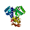
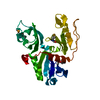
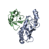
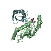
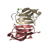
#1: Protein
E3ubiquitin-proteinligaseTRIM69 / RFP-like domain-containing protein trimless / RING finger protein 36 / RING-type E3 ubiquitin ...RFP-like domain-containing protein trimless / RING finger protein 36 / RING-type E3 ubiquitin transferase TRIM69 / Tripartite motif-containing protein 69
Mass: 14741.005 Da / Num. of mol.: 2 Source method: isolated from a genetically manipulated source Source: (gene. exp.) Homo sapiens (human) / Gene: TRIM69, RNF36, HSD-34, HSD34 / Production host: Escherichia coli (E. coli) References: UniProt: Q86WT6, RING-type E3 ubiquitin transferase
Method to determine structure: SAD / Resolution: 2.1→65.46 Å / Cor.coef. Fo:Fc: 0.957 / Cor.coef. Fo:Fc free: 0.937 / SU B: 11.518 / SU ML: 0.142 / SU R Cruickshank DPI: 0.1925 / Cross valid method: THROUGHOUT / σ(F): 0 / ESU R: 0.192 / ESU R Free: 0.172 Details: HYDROGENS HAVE BEEN ADDED IN THE RIDING POSITIONS U VALUES : WITH TLS ADDED
Rfactor
Num. reflection
% reflection
Selection details
Rfree
0.2395
643
5.2 %
RANDOM
Rwork
0.1977
-
-
-
obs
0.1999
11743
99.98 %
-
Solvent computation
Ion probe radii: 0.8 Å / Shrinkage radii: 0.8 Å / VDW probe radii: 1.2 Å
In the structure databanks used in Yorodumi, some data are registered as the other names, "COVID-19 virus" and "2019-nCoV". Here are the details of the virus and the list of structure data.
Jan 31, 2019. EMDB accession codes are about to change! (news from PDBe EMDB page)
EMDB accession codes are about to change! (news from PDBe EMDB page)
The allocation of 4 digits for EMDB accession codes will soon come to an end. Whilst these codes will remain in use, new EMDB accession codes will include an additional digit and will expand incrementally as the available range of codes is exhausted. The current 4-digit format prefixed with “EMD-” (i.e. EMD-XXXX) will advance to a 5-digit format (i.e. EMD-XXXXX), and so on. It is currently estimated that the 4-digit codes will be depleted around Spring 2019, at which point the 5-digit format will come into force.
The EM Navigator/Yorodumi systems omit the EMD- prefix.
Related info.:Q: What is EMD? / ID/Accession-code notation in Yorodumi/EM Navigator
Yorodumi is a browser for structure data from EMDB, PDB, SASBDB, etc.
This page is also the successor to EM Navigator detail page, and also detail information page/front-end page for Omokage search.
The word "yorodu" (or yorozu) is an old Japanese word meaning "ten thousand". "mi" (miru) is to see.
Related info.:EMDB / PDB / SASBDB / Comparison of 3 databanks / Yorodumi Search / Aug 31, 2016. New EM Navigator & Yorodumi / Yorodumi Papers / Jmol/JSmol / Function and homology information / Changes in new EM Navigator and Yorodumi
 Movie
Movie Controller


 Open data
 Basic information
 Components
 Keywords
 Function and homology information
 Homo sapiens (human)
 X-RAY DIFFRACTION /
  Authors
 New Zealand, 2items
  Citation
 Structure visualization
 Downloads & links
 Other downloads


 PDBj
PDBj

 Assembly
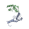


 Mass: 65.409 Da / Num. of mol.: 4 / Source method: obtained synthetically / Formula: Zn
 Mass: 18.015 Da / Num. of mol.: 30 / Source method: isolated from a natural source / Formula: H2O
 Sample preparation
 / Beamline: MX1 / Wavelength: 1.2524 Å
 Processing