 Movie
Movie Controller

[English] 日本語
 Yorodumi
- PDB-6yuq: Capsule O-acetyltransferase of Neisseria meningitidis serogroup A... -
+ Open data

- Basic information
| Entry | Database: PDB / ID: 6yuq | |||||||||
|---|---|---|---|---|---|---|---|---|---|---|
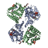
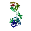
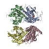
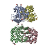
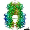
| Title | Capsule O-acetyltransferase of Neisseria meningitidis serogroup A in complex with polysaccharide | |||||||||
 Components Components | SacC | |||||||||
 Keywords Keywords | TRANSFERASE / O-acetyltransferase / a/b hydrolase fold / serine transferase / catalytic triad | |||||||||
| Function / homology |  Function and homology information Function and homology informationUncharacterised protein family (UPF0227) / Alpha/Beta hydrolase fold, catalytic domain / Alpha/Beta hydrolase fold / Rossmann fold / 3-Layer(aba) Sandwich / Alpha Beta Similarity search - Domain/homology | |||||||||
| Biological species |  Neisseria meningitidis serogroup A (bacteria) Neisseria meningitidis serogroup A (bacteria) | |||||||||
| Method |  X-RAY DIFFRACTION / SYNCHROTRON / MOLECULAR REPLACEMENT / Resolution: 1.95 Å X-RAY DIFFRACTION / SYNCHROTRON / MOLECULAR REPLACEMENT / Resolution: 1.95 Å | |||||||||
 Authors Authors | Cramer, J.T. / Fiebig, T. / Fedorov, R. / Muehlenhoff, M. | |||||||||
| Funding support |  Germany, 2items Germany, 2items
| |||||||||
 Citation Citation | Journal: Nat Commun / Year: 2020 Title: Structural and mechanistic basis of capsule O-acetylation in Neisseria meningitidis serogroup A. Authors: Fiebig, T. / Cramer, J.T. / Bethe, A. / Baruch, P. / Curth, U. / Fuhring, J.I. / Buettner, F.F.R. / Vogel, U. / Schubert, M. / Fedorov, R. / Muhlenhoff, M. | |||||||||
| History |
|
- Structure visualization
| Structure viewer | Molecule: MolmilJmol/JSmol |
|---|
- Downloads & links
-Download
| PDBx/mmCIF format | 6yuq.cif.gz | 236.8 KB | Display | PDBx/mmCIF format |
|---|---|---|---|---|
| PDB format | pdb6yuq.ent.gz | 170.8 KB | Display | PDB format |
| PDBx/mmJSON format | 6yuq.json.gz | Tree view | PDBx/mmJSON format | |
| Others |  Other downloads Other downloads |
-Validation report
| Arichive directory | https://data.pdbj.org/pub/pdb/validation_reports/yu/6yuqftp://data.pdbj.org/pub/pdb/validation_reports/yu/6yuq | HTTPS FTP |
|---|
-Related structure data
| Related structure data | 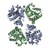 6yuoSC  6yusC 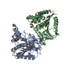 6yuvC S: Starting model for refinement C: citing same article ( |
|---|---|
| Similar structure data |


-Links
 PDBj
PDBj- Assembly
| Deposited unit | 
| ||||||||||||
|---|---|---|---|---|---|---|---|---|---|---|---|---|---|
| 1 | 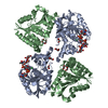
| ||||||||||||
| Unit cell |
|
-Components
-Protein , 1 types, 2 molecules AB
| #1: Protein | Mass: 29374.439 Da / Num. of mol.: 2 Source method: isolated from a genetically manipulated source Source: (gene. exp.) Neisseria meningitidis serogroup A (bacteria)Gene: sacC / Production host: |
|---|
-Sugars , 2 types, 4 molecules 
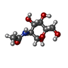
| #5: Sugar |  Type: D-saccharide, alpha linking / Mass: 301.188 Da / Num. of mol.: 3 Type: D-saccharide, alpha linking / Mass: 301.188 Da / Num. of mol.: 3Source method: isolated from a genetically manipulated source Formula: C8H16NO9P / Feature type: SUBJECT OF INVESTIGATION #6: Sugar | ChemComp-BM3 / |  Type: D-saccharide, alpha linking / Mass: 221.208 Da / Num. of mol.: 1 Type: D-saccharide, alpha linking / Mass: 221.208 Da / Num. of mol.: 1Source method: isolated from a genetically manipulated source Formula: C8H15NO6 / Feature type: SUBJECT OF INVESTIGATION |
|---|
-Non-polymers , 5 types, 52 molecules 


| #2: Chemical | ChemComp-EDO /  Mass: 62.068 Da / Num. of mol.: 1 / Source method: obtained synthetically / Formula: C2H6O2 Mass: 62.068 Da / Num. of mol.: 1 / Source method: obtained synthetically / Formula: C2H6O2 | ||||
|---|---|---|---|---|---|
| #3: Chemical | ChemComp-PEG /  Mass: 106.120 Da / Num. of mol.: 1 / Source method: obtained synthetically / Formula: C4H10O3 Mass: 106.120 Da / Num. of mol.: 1 / Source method: obtained synthetically / Formula: C4H10O3 | ||||
| #4: Chemical |  Mass: 35.453 Da / Num. of mol.: 2 / Source method: obtained synthetically / Formula: Cl / Feature type: SUBJECT OF INVESTIGATION Mass: 35.453 Da / Num. of mol.: 2 / Source method: obtained synthetically / Formula: Cl / Feature type: SUBJECT OF INVESTIGATION#7: Chemical | ChemComp-PGE / |  Mass: 150.173 Da / Num. of mol.: 1 Mass: 150.173 Da / Num. of mol.: 1Source method: isolated from a genetically manipulated source Formula: C6H14O4 #8: Water | ChemComp-HOH / | Mass: 18.015 Da / Num. of mol.: 47 / Source method: isolated from a natural source / Formula: H2O |
-Details
| Has ligand of interest | Y |
|---|
-Experimental details
-Experiment
| Experiment | Method: X-RAY DIFFRACTION / Number of used crystals: 1 |
|---|
- Sample preparation
| Crystal | Density Matthews: 2.83 Å3/Da / Density % sol: 56.47 % |
|---|---|
| Crystal grow | Temperature: 291.15 K / Method: vapor diffusion, sitting drop Details: Native wild type CsaC crystallized in sitting drop setups at concentrations of approx. 18mg/ml. Fine screens around initial screening conditions resulted in many isomorphous crystals. Mother ...Details: Native wild type CsaC crystallized in sitting drop setups at concentrations of approx. 18mg/ml. Fine screens around initial screening conditions resulted in many isomorphous crystals. Mother liquor contained 50mM HEPES pH 7.0, 100 mM HEPES pH 7.6, 100mM NaCl, 5mM MgCl2, 1mM EDTA, and 31-42% PEG200. Good quality crystals grew at 4, 12, and 18C. PH range: 7.6 |
-Data collection
| Diffraction | Mean temperature: 100 K / Serial crystal experiment: N | |||||||||||||||||||||||||||||||||||||||||||||||||||||||||||||||||||||||||||||||||||||||||||||||||||
|---|---|---|---|---|---|---|---|---|---|---|---|---|---|---|---|---|---|---|---|---|---|---|---|---|---|---|---|---|---|---|---|---|---|---|---|---|---|---|---|---|---|---|---|---|---|---|---|---|---|---|---|---|---|---|---|---|---|---|---|---|---|---|---|---|---|---|---|---|---|---|---|---|---|---|---|---|---|---|---|---|---|---|---|---|---|---|---|---|---|---|---|---|---|---|---|---|---|---|---|---|
| Diffraction source | Source: SYNCHROTRON / Site: PETRA III, EMBL c/o DESY / Beamline: P14 (MX2) / Wavelength: 0.97625 Å | |||||||||||||||||||||||||||||||||||||||||||||||||||||||||||||||||||||||||||||||||||||||||||||||||||
| Detector | Type: DECTRIS EIGER X 16M / Detector: PIXEL / Date: Sep 24, 2017 | |||||||||||||||||||||||||||||||||||||||||||||||||||||||||||||||||||||||||||||||||||||||||||||||||||
| Radiation | Protocol: SINGLE WAVELENGTH / Monochromatic (M) / Laue (L): M / Scattering type: x-ray | |||||||||||||||||||||||||||||||||||||||||||||||||||||||||||||||||||||||||||||||||||||||||||||||||||
| Radiation wavelength | Wavelength: 0.97625 Å / Relative weight: 1 | |||||||||||||||||||||||||||||||||||||||||||||||||||||||||||||||||||||||||||||||||||||||||||||||||||
| Reflection | Resolution: 1.95→48.62 Å / Num. obs: 49502 / % possible obs: 99.9 % / Redundancy: 26.2 % / CC1/2: 0.999 / Rmerge(I) obs: 0.059 / Rpim(I) all: 0.012 / Rrim(I) all: 0.06 / Net I/σ(I): 25.1 | |||||||||||||||||||||||||||||||||||||||||||||||||||||||||||||||||||||||||||||||||||||||||||||||||||
| Reflection shell | Diffraction-ID: 1
|
- Processing
| Software |
| |||||||||||||||||||||||||||||||||||||||||||||||||||||||||||||||||||||||||||||||||||||||||||||||||||||||||||||||||||||||||||||||||||||
|---|---|---|---|---|---|---|---|---|---|---|---|---|---|---|---|---|---|---|---|---|---|---|---|---|---|---|---|---|---|---|---|---|---|---|---|---|---|---|---|---|---|---|---|---|---|---|---|---|---|---|---|---|---|---|---|---|---|---|---|---|---|---|---|---|---|---|---|---|---|---|---|---|---|---|---|---|---|---|---|---|---|---|---|---|---|---|---|---|---|---|---|---|---|---|---|---|---|---|---|---|---|---|---|---|---|---|---|---|---|---|---|---|---|---|---|---|---|---|---|---|---|---|---|---|---|---|---|---|---|---|---|---|---|---|
| Refinement | Method to determine structure: MOLECULAR REPLACEMENT Starting model: 6YUO Resolution: 1.95→46.27 Å / SU ML: 0.2771 / Cross valid method: FREE R-VALUE / σ(F): 1.34 / Phase error: 29.6893
| |||||||||||||||||||||||||||||||||||||||||||||||||||||||||||||||||||||||||||||||||||||||||||||||||||||||||||||||||||||||||||||||||||||
| Solvent computation | Shrinkage radii: 0.9 Å / VDW probe radii: 1.11 Å | |||||||||||||||||||||||||||||||||||||||||||||||||||||||||||||||||||||||||||||||||||||||||||||||||||||||||||||||||||||||||||||||||||||
| Displacement parameters | Biso mean: 71.33 Å2 | |||||||||||||||||||||||||||||||||||||||||||||||||||||||||||||||||||||||||||||||||||||||||||||||||||||||||||||||||||||||||||||||||||||
| Refinement step | Cycle: LAST / Resolution: 1.95→46.27 Å
| |||||||||||||||||||||||||||||||||||||||||||||||||||||||||||||||||||||||||||||||||||||||||||||||||||||||||||||||||||||||||||||||||||||
| Refine LS restraints |
| |||||||||||||||||||||||||||||||||||||||||||||||||||||||||||||||||||||||||||||||||||||||||||||||||||||||||||||||||||||||||||||||||||||
| LS refinement shell |
| |||||||||||||||||||||||||||||||||||||||||||||||||||||||||||||||||||||||||||||||||||||||||||||||||||||||||||||||||||||||||||||||||||||
| Refinement TLS params. | Method: refined / Refine-ID: X-RAY DIFFRACTION
| |||||||||||||||||||||||||||||||||||||||||||||||||||||||||||||||||||||||||||||||||||||||||||||||||||||||||||||||||||||||||||||||||||||
| Refinement TLS group | Refine-ID: X-RAY DIFFRACTION / Auth seq-ID: 1 - 245 / Label seq-ID: 1 - 245
|
