 Movie
Movie Controller


+ Open data

- Basic information
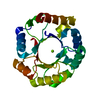
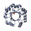
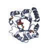
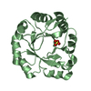
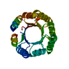
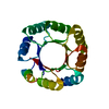
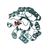
| Entry | Database: PDB / ID: 6yqx | |||||||||
|---|---|---|---|---|---|---|---|---|---|---|
| Title | Crystal structure of DeNovoTIM13, a de novo designed TIM barrel | |||||||||
 Components Components | de novo designed TIM barrel DeNovoTIM13 | |||||||||
 Keywords Keywords | DE NOVO PROTEIN / de novo protein design / epistasis / stability landscape / TIM barrel / (beta/alfa)8 barrel | |||||||||
| Biological species | synthetic construct (others) | |||||||||
| Method |  X-RAY DIFFRACTION / SYNCHROTRON / MOLECULAR REPLACEMENT / Resolution: 1.638 Å X-RAY DIFFRACTION / SYNCHROTRON / MOLECULAR REPLACEMENT / Resolution: 1.638 Å | |||||||||
 Authors Authors | Romero-Romero, S. / Kordes, S. / Shanmugaratnam, S. / Fernandez-Velasco, D.A. / Hocker, B. | |||||||||
| Funding support |  Mexico, 2items Mexico, 2items
| |||||||||
 Citation Citation | Journal: J.Mol.Biol. / Year: 2021 Title: The Stability Landscape of de novo TIM Barrels Explored by a Modular Design Approach. Authors: Romero-Romero, S. / Costas, M. / Silva Manzano, D.A. / Kordes, S. / Rojas-Ortega, E. / Tapia, C. / Guerra, Y. / Shanmugaratnam, S. / Rodriguez-Romero, A. / Baker, D. / Hocker, B. / Fernandez-Velasco, D.A. | |||||||||
| History |
|
- Structure visualization
| Structure viewer | Molecule:  MolmilJmol/JSmol MolmilJmol/JSmol |
|---|
- Downloads & links
-Download
| PDBx/mmCIF format | 6yqx.cif.gz | 89.8 KB | Display | PDBx/mmCIF format |
|---|---|---|---|---|
| PDB format | pdb6yqx.ent.gz | 67.7 KB | Display | PDB format |
| PDBx/mmJSON format | 6yqx.json.gz | Tree view | PDBx/mmJSON format | |
| Others |  Other downloads Other downloads |
-Validation report
| Arichive directory | https://data.pdbj.org/pub/pdb/validation_reports/yq/6yqxftp://data.pdbj.org/pub/pdb/validation_reports/yq/6yqx | HTTPS FTP |
|---|
-Related structure data


-Links
 PDBj
PDBj
- Assembly
| Deposited unit | 
| ||||||||
|---|---|---|---|---|---|---|---|---|---|
| 1 |
| ||||||||
| Unit cell |
|
-Components
| #1: Protein | Mass: 22117.680 Da / Num. of mol.: 1 Source method: isolated from a genetically manipulated source Details: M (N-terminal) and GLEHHHHHH (C-terminal) come from the expresion vector. Source: (gene. exp.) synthetic construct (others) / Plasmid: pET29b(+) / Production host:  |
|---|---|
| #2: Chemical | ChemComp-GOL /   Mass: 92.094 Da / Num. of mol.: 1 / Source method: obtained synthetically / Formula: C3H8O3 Mass: 92.094 Da / Num. of mol.: 1 / Source method: obtained synthetically / Formula: C3H8O3 |
| #3: Chemical | ChemComp-CL /   Mass: 35.453 Da / Num. of mol.: 1 / Source method: obtained synthetically / Formula: Cl Mass: 35.453 Da / Num. of mol.: 1 / Source method: obtained synthetically / Formula: Cl |
| #4: Water | ChemComp-HOH /  Mass: 18.015 Da / Num. of mol.: 128 / Source method: isolated from a natural source / Formula: H2O Mass: 18.015 Da / Num. of mol.: 128 / Source method: isolated from a natural source / Formula: H2O |
| Has ligand of interest | N |
-Experimental details
-Experiment
| Experiment | Method: X-RAY DIFFRACTION / Number of used crystals: 1 |
|---|
- Sample preparation
| Crystal | Density Matthews: 2.23 Å3/Da / Density % sol: 44.93 % |
|---|---|
| Crystal grow | Temperature: 293.15 K / Method: vapor diffusion, sitting drop / pH: 8.5 Details: 0.17 M sodium acetate, 0.085 M TRIS pH: 8.5, 25% w/v PEG 4000, 15% v/v Glycerol. |
-Data collection
| Diffraction | Mean temperature: 100 K / Serial crystal experiment: N |
|---|---|
| Diffraction source | Source: SYNCHROTRON / Site: SLS  / Beamline: X06DA / Wavelength: 1 Å / Beamline: X06DA / Wavelength: 1 Å |
| Detector | Type: DECTRIS PILATUS 2M-F / Detector: PIXEL / Date: Dec 11, 2016 |
| Radiation | Monochromator: M / Protocol: SINGLE WAVELENGTH / Monochromatic (M) / Laue (L): M / Scattering type: x-ray |
| Radiation wavelength | Wavelength: 1 Å / Relative weight: 1 |
| Reflection | Resolution: 1.638→44.793 Å / Num. obs: 23407 / % possible obs: 99.5 % / Redundancy: 3.4 % / Biso Wilson estimate: 28.4 Å2 / CC1/2: 0.998 / Rmerge(I) obs: 0.042 / Net I/σ(I): 14.3 |
| Reflection shell | Resolution: 1.638→1.7 Å / Redundancy: 3.2 % / Rmerge(I) obs: 0.552 / Mean I/σ(I) obs: 1.5 / Num. unique obs: 2280 / CC1/2: 0.659 / % possible all: 97.6 |
- Processing
| Software |
| ||||||||||||||||||||||||||||||||||||||||||||||||||||||||||||||||||||||||||||||||||||||||||
|---|---|---|---|---|---|---|---|---|---|---|---|---|---|---|---|---|---|---|---|---|---|---|---|---|---|---|---|---|---|---|---|---|---|---|---|---|---|---|---|---|---|---|---|---|---|---|---|---|---|---|---|---|---|---|---|---|---|---|---|---|---|---|---|---|---|---|---|---|---|---|---|---|---|---|---|---|---|---|---|---|---|---|---|---|---|---|---|---|---|---|---|
| Refinement | Method to determine structure: MOLECULAR REPLACEMENT Starting model: Rosetta backbone model Resolution: 1.638→44.793 Å / SU ML: 0.22 / Cross valid method: THROUGHOUT / σ(F): 1.99 / Phase error: 20.68
| ||||||||||||||||||||||||||||||||||||||||||||||||||||||||||||||||||||||||||||||||||||||||||
| Solvent computation | Shrinkage radii: 0.9 Å / VDW probe radii: 1.11 Å | ||||||||||||||||||||||||||||||||||||||||||||||||||||||||||||||||||||||||||||||||||||||||||
| Displacement parameters | Biso max: 99.67 Å2 / Biso mean: 39.2143 Å2 / Biso min: 16.68 Å2 | ||||||||||||||||||||||||||||||||||||||||||||||||||||||||||||||||||||||||||||||||||||||||||
| Refinement step | Cycle: final / Resolution: 1.638→44.793 Å
| ||||||||||||||||||||||||||||||||||||||||||||||||||||||||||||||||||||||||||||||||||||||||||
| LS refinement shell | Refine-ID: X-RAY DIFFRACTION / Rfactor Rfree error: 0
| ||||||||||||||||||||||||||||||||||||||||||||||||||||||||||||||||||||||||||||||||||||||||||
| Refinement TLS params. | Method: refined / Origin x: 4.2974 Å / Origin y: 16.4939 Å / Origin z: -2.6164 Å
| ||||||||||||||||||||||||||||||||||||||||||||||||||||||||||||||||||||||||||||||||||||||||||
| Refinement TLS group | Selection details: (chain A and resseq 2:184) |
