 Movie
Movie Controller

[English] 日本語
 Yorodumi
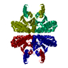
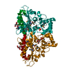
- PDB-6yjn: Crystal structure of beta carbonic anhydrase from the pathogenic ... -
+ Open data

- Basic information
| Entry | Database: PDB / ID: 6yjn | ||||||
|---|---|---|---|---|---|---|---|
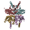
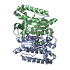
| Title | Crystal structure of beta carbonic anhydrase from the pathogenic bacterium Burkholderia pseudomallei. | ||||||
 Components Components | Beta carbonic anhydrase | ||||||
 Keywords Keywords | LYASE / beta carbonic anhydrase / Burkholderia pseudomallei | ||||||
| Function / homology |  Function and homology information Function and homology informationcarbon utilization / carbonic anhydrase / carbonate dehydratase activity / zinc ion binding Similarity search - Function | ||||||
| Biological species |  Burkholderia pseudomallei (bacteria) Burkholderia pseudomallei (bacteria) | ||||||
| Method |  X-RAY DIFFRACTION / SYNCHROTRON / MOLECULAR REPLACEMENT / Resolution: 2.7 Å X-RAY DIFFRACTION / SYNCHROTRON / MOLECULAR REPLACEMENT / Resolution: 2.7 Å | ||||||
 Authors Authors | Angeli, A. / Ferraroni, M. | ||||||
 Citation Citation | Journal: Molecules / Year: 2020 Title: Crystal Structure of a Tetrameric Type II beta-Carbonic Anhydrase from the Pathogenic BacteriumBurkholderia pseudomallei. Authors: Angeli, A. / Ferraroni, M. / Pinteala, M. / Maier, S.S. / Simionescu, B.C. / Carta, F. / Del Prete, S. / Capasso, C. / Supuran, C.T. | ||||||
| History |
|
- Structure visualization
| Structure viewer | Molecule: MolmilJmol/JSmol |
|---|
- Downloads & links
-Download
| PDBx/mmCIF format | 6yjn.cif.gz | 57.1 KB | Display | PDBx/mmCIF format |
|---|---|---|---|---|
| PDB format | pdb6yjn.ent.gz | 39.2 KB | Display | PDB format |
| PDBx/mmJSON format | 6yjn.json.gz | Tree view | PDBx/mmJSON format | |
| Others |  Other downloads Other downloads |
-Validation report
| Arichive directory | https://data.pdbj.org/pub/pdb/validation_reports/yj/6yjnftp://data.pdbj.org/pub/pdb/validation_reports/yj/6yjn | HTTPS FTP |
|---|
-Related structure data
| Related structure data | 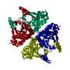 6yl7C 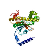 4rxyS S: Starting model for refinement C: citing same article ( |
|---|---|
| Similar structure data |


-Links
 PDBj
PDBj
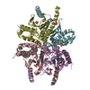
- Assembly
| Deposited unit | 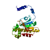
| ||||||||||||||||||
|---|---|---|---|---|---|---|---|---|---|---|---|---|---|---|---|---|---|---|---|
| 1 | 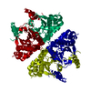
| ||||||||||||||||||
| Unit cell |
| ||||||||||||||||||
| Components on special symmetry positions |
|
-Components
| #1: Protein | Mass: 28269.016 Da / Num. of mol.: 1 Source method: isolated from a genetically manipulated source Source: (gene. exp.) Burkholderia pseudomallei (bacteria)Gene: can_1, can_2, BOC42_16450, CXQ84_19645, DF122_01465, ERS013345_01543, SAMEA1968934_03740 Production host: References: UniProt: A0A069AXA0, UniProt: Q63Y17*PLUS, carbonic anhydrase |
|---|---|
| #2: Chemical | ChemComp-ZN /   Mass: 65.409 Da / Num. of mol.: 1 / Source method: obtained synthetically / Formula: Zn / Feature type: SUBJECT OF INVESTIGATION Mass: 65.409 Da / Num. of mol.: 1 / Source method: obtained synthetically / Formula: Zn / Feature type: SUBJECT OF INVESTIGATION |
| #3: Water | ChemComp-HOH /  Mass: 18.015 Da / Num. of mol.: 52 / Source method: isolated from a natural source / Formula: H2O Mass: 18.015 Da / Num. of mol.: 52 / Source method: isolated from a natural source / Formula: H2O |
| Has ligand of interest | Y |
-Experimental details
-Experiment
| Experiment | Method: X-RAY DIFFRACTION / Number of used crystals: 1 |
|---|
- Sample preparation
| Crystal | Density Matthews: 2.21 Å3/Da / Density % sol: 44.31 % |
|---|---|
| Crystal grow | Temperature: 293 K / Method: vapor diffusion, sitting drop / pH: 8.5 Details: 22% PEG 4000, 10% isopropanol, 100 mM HEPES pH 8.5 and 3% v/v 1,5-Diaminopentene di-HCl. |
-Data collection
| Diffraction | Mean temperature: 100 K / Serial crystal experiment: N |
|---|---|
| Diffraction source | Source: SYNCHROTRON / Site: ELETTRA  / Beamline: 11.2C / Wavelength: 1 Å / Beamline: 11.2C / Wavelength: 1 Å |
| Detector | Type: DECTRIS PILATUS3 S 6M / Detector: PIXEL / Date: Sep 16, 2019 |
| Radiation | Protocol: SINGLE WAVELENGTH / Monochromatic (M) / Laue (L): M / Scattering type: x-ray |
| Radiation wavelength | Wavelength: 1 Å / Relative weight: 1 |
| Reflection | Resolution: 2.7→45.49 Å / Num. obs: 7508 / % possible obs: 99.9 % / Redundancy: 32.7 % / CC1/2: 0.999 / Rmerge(I) obs: 0.28 / Rpim(I) all: 0.067 / Rrim(I) all: 0.288 / Net I/σ(I): 12.6 |
| Reflection shell | Resolution: 2.7→2.83 Å / Rmerge(I) obs: 8.711 / Num. unique obs: 968 / CC1/2: 0.415 / Rpim(I) all: 2.059 / Rrim(I) all: 8.953 |
- Processing
| Software |
| |||||||||||||||||||||||||||||||||||||||||||||
|---|---|---|---|---|---|---|---|---|---|---|---|---|---|---|---|---|---|---|---|---|---|---|---|---|---|---|---|---|---|---|---|---|---|---|---|---|---|---|---|---|---|---|---|---|---|---|
| Refinement | Method to determine structure: MOLECULAR REPLACEMENT Starting model: 4rxy Resolution: 2.7→45.08 Å / Cor.coef. Fo:Fc: 0.96 / Cor.coef. Fo:Fc free: 0.894 / SU B: 22.437 / SU ML: 0.424 / Cross valid method: THROUGHOUT / σ(F): 0 / ESU R: 0.859 / ESU R Free: 0.419 / Stereochemistry target values: MAXIMUM LIKELIHOOD / Details: U VALUES : REFINED INDIVIDUALLY
| |||||||||||||||||||||||||||||||||||||||||||||
| Solvent computation | Ion probe radii: 0.8 Å / Shrinkage radii: 0.8 Å / VDW probe radii: 1.2 Å / Solvent model: MASK | |||||||||||||||||||||||||||||||||||||||||||||
| Displacement parameters | Biso max: 202.03 Å2 / Biso mean: 81.594 Å2 / Biso min: 49.03 Å2
| |||||||||||||||||||||||||||||||||||||||||||||
| Refinement step | Cycle: final / Resolution: 2.7→45.08 Å
| |||||||||||||||||||||||||||||||||||||||||||||
| Refine LS restraints |
| |||||||||||||||||||||||||||||||||||||||||||||
| LS refinement shell | Resolution: 2.701→2.771 Å / Rfactor Rfree error: 0 / Total num. of bins used: 20
|
