 Movie
Movie Controller

[English] 日本語
 Yorodumi
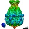
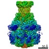
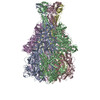
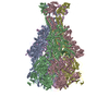
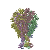
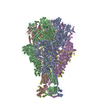
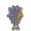
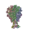
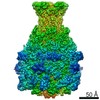
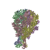
- PDB-6yey: Xenorhabdus nematophila XptA1 in complex with porcine mucosa heparin -
+ Open data

- Basic information
| Entry | Database: PDB / ID: 6yey | ||||||
|---|---|---|---|---|---|---|---|
| Title | Xenorhabdus nematophila XptA1 in complex with porcine mucosa heparin | ||||||
 Components Components | A component of insecticidal toxin complex (Tc) | ||||||
 Keywords Keywords | TOXIN / complex / glycan | ||||||
| Function / homology | TcA receptor binding domain / TcA receptor binding domain / Insecticidal toxin complex/plasmid virulence protein / Tc toxin complex TcA, C-terminal TcB-binding domain / Neuraminidase-like domain / Salmonella virulence plasmid 28.1kDa A protein / Tc toxin complex TcA C-terminal TcB-binding domain / Neuraminidase-like domain / A component of insecticidal toxin complex (Tc) Function and homology information Function and homology information | ||||||
| Biological species |  Xenorhabdus nematophila (bacteria) Xenorhabdus nematophila (bacteria) | ||||||
| Method | ELECTRON MICROSCOPY / single particle reconstruction / cryo EM / Resolution: 3.7 Å | ||||||
 Authors Authors | Roderer, D. / Broecker, F. / Sitsel, O. / Kaplonek, P. / Leidreiter, F. / Seeberger, P.H. / Raunser, S. | ||||||
| Funding support |  Germany, 1items Germany, 1items
| ||||||
 Citation Citation | Journal: Nat Commun / Year: 2020 Title: Glycan-dependent cell adhesion mechanism of Tc toxins. Authors: Daniel Roderer / Felix Bröcker / Oleg Sitsel / Paulina Kaplonek / Franziska Leidreiter / Peter H Seeberger / Stefan Raunser /  Abstract: Toxin complex (Tc) toxins are virulence factors of pathogenic bacteria. Tcs are composed of three subunits: TcA, TcB and TcC. TcA facilitates receptor-toxin interaction and membrane permeation, TcB ...Toxin complex (Tc) toxins are virulence factors of pathogenic bacteria. Tcs are composed of three subunits: TcA, TcB and TcC. TcA facilitates receptor-toxin interaction and membrane permeation, TcB and TcC form a toxin-encapsulating cocoon. While the mechanisms of holotoxin assembly and pore formation have been described, little is known about receptor binding of TcAs. Here, we identify heparins/heparan sulfates and Lewis antigens as receptors for different TcAs from insect and human pathogens. Glycan array screening reveals that all tested TcAs bind negatively charged heparins. Cryo-EM structures of Morganella morganii TcdA4 and Xenorhabdus nematophila XptA1 reveal that heparins/heparan sulfates unexpectedly bind to different regions of the shell domain, including receptor-binding domains. In addition, Photorhabdus luminescens TcdA1 binds to Lewis antigens with micromolar affinity. Here, the glycan interacts with the receptor-binding domain D of the toxin. Our results suggest a glycan dependent association mechanism of Tc toxins on the host cell surface. | ||||||
| History |
|
- Structure visualization
| Movie |
Movie viewer |
|---|---|
| Structure viewer | Molecule: MolmilJmol/JSmol |
- Downloads & links
-Download
| PDBx/mmCIF format | 6yey.cif.gz | 1.9 MB | Display | PDBx/mmCIF format |
|---|---|---|---|---|
| PDB format | pdb6yey.ent.gz | 1.6 MB | Display | PDB format |
| PDBx/mmJSON format | 6yey.json.gz | Tree view | PDBx/mmJSON format | |
| Others |  Other downloads Other downloads |
-Validation report
| Arichive directory | https://data.pdbj.org/pub/pdb/validation_reports/ye/6yeyftp://data.pdbj.org/pub/pdb/validation_reports/ye/6yey | HTTPS FTP |
|---|
-Related structure data
| Related structure data | 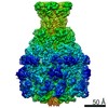 10797MC 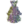 6yewC M: map data used to model this data C: citing same article ( |
|---|---|
| Similar structure data |

-Links
 PDBj
PDBj- Assembly
| Deposited unit | 
|
|---|---|
| 1 |
|
-Components
| #1: Protein | Mass: 287120.781 Da / Num. of mol.: 5 Source method: isolated from a genetically manipulated source Source: (gene. exp.) Xenorhabdus nematophila (bacteria) / Gene: xptA, XNC2_2467 / Production host: |
|---|
-Experimental details
-Experiment
| Experiment | Method: ELECTRON MICROSCOPY |
|---|---|
| EM experiment | Aggregation state: PARTICLE / 3D reconstruction method: single particle reconstruction |
- Sample preparation
| Component | Name: Xenorhabdus nematophila XptA1 pentamer in complex with porcine mucosa heparin Type: COMPLEX / Entity ID: all / Source: RECOMBINANT |
|---|---|
| Molecular weight | Value: 1.4 MDa / Experimental value: NO |
| Source (natural) | Organism: Xenorhabdus nematophila (bacteria) |
| Source (recombinant) | Organism: |
| Buffer solution | pH: 7.5 |
| Specimen | Conc.: 0.1 mg/ml / Embedding applied: NO / Shadowing applied: NO / Staining applied: NO / Vitrification applied: YES Details: 0.10 mg/ml XptA1 were pre-applied on the grid for 30s. Subsequently, the grid was manually blotted and 0.30 mg/ml porcine mucosa heparin was applied and incubated for 4 min. |
| Specimen support | Grid material: GOLD / Grid type: Quantifoil R2/1 |
| Vitrification | Instrument: FEI VITROBOT MARK IV / Cryogen name: ETHANE / Humidity: 100 % / Chamber temperature: 285 K |
- Electron microscopy imaging
| Experimental equipment |  Model: Talos Arctica / Image courtesy: FEI Company |
|---|---|
| Microscopy | Model: FEI TALOS ARCTICA |
| Electron gun | Electron source:  FIELD EMISSION GUN / Accelerating voltage: 200 kV / Illumination mode: SPOT SCAN FIELD EMISSION GUN / Accelerating voltage: 200 kV / Illumination mode: SPOT SCAN |
| Electron lens | Mode: BRIGHT FIELD / Alignment procedure: COMA FREE |
| Image recording | Electron dose: 52 e/Å2 / Detector mode: INTEGRATING / Film or detector model: FEI FALCON III (4k x 4k) / Num. of real images: 1855 |
- Processing
| Software | Name: PHENIX / Version: 1.15.2_3472: / Classification: refinement | ||||||||||||||||||||||||||||||||
|---|---|---|---|---|---|---|---|---|---|---|---|---|---|---|---|---|---|---|---|---|---|---|---|---|---|---|---|---|---|---|---|---|---|
| EM software |
| ||||||||||||||||||||||||||||||||
| CTF correction | Type: PHASE FLIPPING AND AMPLITUDE CORRECTION | ||||||||||||||||||||||||||||||||
| Particle selection | Num. of particles selected: 346048 | ||||||||||||||||||||||||||||||||
| Symmetry | Point symmetry: C5 (5 fold cyclic) | ||||||||||||||||||||||||||||||||
| 3D reconstruction | Resolution: 3.7 Å / Resolution method: FSC 0.143 CUT-OFF / Num. of particles: 172596 / Symmetry type: POINT | ||||||||||||||||||||||||||||||||
| Atomic model building | B value: 65.8 / Protocol: FLEXIBLE FIT / Space: REAL | ||||||||||||||||||||||||||||||||
| Atomic model building | PDB-ID: 6RW8 Accession code: 6RW8 / Source name: PDB / Type: experimental model | ||||||||||||||||||||||||||||||||
| Refine LS restraints |
|
