 Movie
Movie Controller


+ Open data

- Basic information
| Entry | Database: PDB / ID: 6rw6 | ||||||
|---|---|---|---|---|---|---|---|
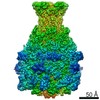
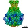
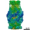
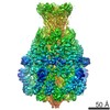
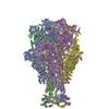
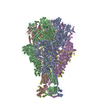
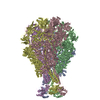
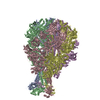
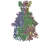
| Title | Cryo-EM structure of Photorhabdus luminescens TcdA1 | ||||||
 Components Components | TcdA1 | ||||||
 Keywords Keywords | TOXIN / membrane permeation / translocation / complex | ||||||
| Function / homology |  Function and homology information Function and homology information | ||||||
| Biological species |  Photorhabdus luminescens (bacteria) Photorhabdus luminescens (bacteria) | ||||||
| Method | ELECTRON MICROSCOPY / single particle reconstruction / cryo EM / Resolution: 2.75 Å | ||||||
 Authors Authors | Roderer, D. / Leidreiter, F. / Gatsogiannis, C. / Meusch, D. / Benz, R. / Raunser, S. | ||||||
| Funding support |  Germany, 1items Germany, 1items
| ||||||
 Citation Citation | Journal: Sci Adv / Year: 2019 Title: Common architecture of Tc toxins from human and insect pathogenic bacteria. Authors: F Leidreiter / D Roderer / D Meusch / C Gatsogiannis / R Benz / S Raunser / Abstract: Tc toxins use a syringe-like mechanism to penetrate the membrane and translocate toxic enzymes into the host cytosol. They are composed of three components: TcA, TcB, and TcC. Low-resolution ...Tc toxins use a syringe-like mechanism to penetrate the membrane and translocate toxic enzymes into the host cytosol. They are composed of three components: TcA, TcB, and TcC. Low-resolution structures of TcAs from different bacteria suggest a considerable difference in their architecture and possibly in their mechanism of action. Here, we present high-resolution structures of five TcAs from insect and human pathogens, which show a similar overall composition and domain organization. Essential structural features, including a trefoil protein knot, are present in all TcAs, suggesting a common mechanism of action. All TcAs form functional pores and can be combined with TcB-TcC subunits from other species to form active chimeric holotoxins. We identified a conserved ionic pair that stabilizes the shell, likely operating as a strong latch that only springs open after destabilization of other regions. Our results provide new insights into the architecture and mechanism of the Tc toxin family. | ||||||
| History |
|
- Structure visualization
| Movie |
Movie viewer |
|---|---|
| Structure viewer | Molecule: MolmilJmol/JSmol |
- Downloads & links
-Download
| PDBx/mmCIF format | 6rw6.cif.gz | 2.3 MB | Display | PDBx/mmCIF format |
|---|---|---|---|---|
| PDB format | pdb6rw6.ent.gz | 1.9 MB | Display | PDB format |
| PDBx/mmJSON format | 6rw6.json.gz | Tree view | PDBx/mmJSON format | |
| Others |  Other downloads Other downloads |
-Validation report
| Arichive directory | https://data.pdbj.org/pub/pdb/validation_reports/rw/6rw6ftp://data.pdbj.org/pub/pdb/validation_reports/rw/6rw6 | HTTPS FTP |
|---|
-Related structure data
| Related structure data | 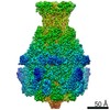 10033MC 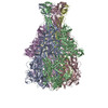 6rw8C 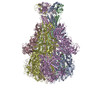 6rw9C 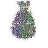 6rwaC 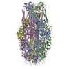 6rwbC M: map data used to model this data C: citing same article ( |
|---|---|
| Similar structure data |

-Links
 PDBj
PDBj- Assembly
| Deposited unit | 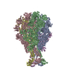
|
|---|---|
| 1 |
|
-Components
| #1: Protein | Mass: 283229.406 Da / Num. of mol.: 5 Source method: isolated from a genetically manipulated source Source: (gene. exp.) Photorhabdus luminescens (bacteria) / Gene: tcdA, tcdA1 / Production host: |
|---|
-Experimental details
-Experiment
| Experiment | Method: ELECTRON MICROSCOPY |
|---|---|
| EM experiment | Aggregation state: PARTICLE / 3D reconstruction method: single particle reconstruction |
- Sample preparation
| Component | Name: TcdA1 pentamer / Type: COMPLEX / Entity ID: all / Source: RECOMBINANT |
|---|---|
| Molecular weight | Value: 1.4 MDa / Experimental value: NO |
| Source (natural) | Organism: Photorhabdus luminescens (bacteria) |
| Source (recombinant) | Organism: |
| Buffer solution | pH: 8 |
| Specimen | Embedding applied: NO / Shadowing applied: NO / Staining applied: NO / Vitrification applied: YES |
| Vitrification | Cryogen name: ETHANE |
- Electron microscopy imaging
| Experimental equipment |  Model: Titan Krios / Image courtesy: FEI Company |
|---|---|
| Microscopy | Model: FEI TITAN KRIOS |
| Electron gun | Electron source:  FIELD EMISSION GUN / Accelerating voltage: 300 kV / Illumination mode: SPOT SCAN FIELD EMISSION GUN / Accelerating voltage: 300 kV / Illumination mode: SPOT SCAN |
| Electron lens | Mode: BRIGHT FIELD |
| Image recording | Electron dose: 100 e/Å2 / Film or detector model: FEI FALCON II (4k x 4k) |
- Processing
| EM software |
| ||||||||||||||||||
|---|---|---|---|---|---|---|---|---|---|---|---|---|---|---|---|---|---|---|---|
| CTF correction | Type: PHASE FLIPPING AND AMPLITUDE CORRECTION | ||||||||||||||||||
| Symmetry | Point symmetry: C5 (5 fold cyclic) | ||||||||||||||||||
| 3D reconstruction | Resolution: 2.75 Å / Resolution method: FSC 0.143 CUT-OFF / Num. of particles: 436273 / Symmetry type: POINT | ||||||||||||||||||
| Atomic model building | PDB-ID: 1VW1 1vw1 Accession code: 1VW1 / Source name: PDB / Type: experimental model |
