 Movie
Movie Controller

[English] 日本語
 Yorodumi
- PDB-6yeg: Hybrid structure of the SPP1 tail tube by solid-state NMR and cry... -
+ Open data

- Basic information
| Entry | Database: PDB / ID: 6yeg | ||||||||||||||||||||||||||||
|---|---|---|---|---|---|---|---|---|---|---|---|---|---|---|---|---|---|---|---|---|---|---|---|---|---|---|---|---|---|
| Title | Hybrid structure of the SPP1 tail tube by solid-state NMR and cryo EM - Final EM Refinement | ||||||||||||||||||||||||||||
 Components Components | Tail tube protein gp17.1* Keywords KeywordsVIRAL PROTEIN / complex / tail tube / scaffolding / DNA transport | Function / homology |  Function and homology information Function and homology informationviral head-tail joining / virus tail, tube / symbiont genome ejection through host cell envelope, long flexible tail mechanism / viral translational frameshifting Similarity search - Function Biological species |  Bacillus phage SPP1 (virus) Bacillus phage SPP1 (virus)Method | ELECTRON MICROSCOPY / SOLID-STATE NMR / helical reconstruction / na / cryo EM / Resolution: 4 Å |  Authors AuthorsZinke, M. / Sachowsky, K.A.A. / Zinn-Justin, S. / Ravelli, R. / Schroder, G.F. / Habeck, M. / Lange, A. | Funding support | European Union, 1items |
 CitationJournal: Nat Commun / Year: 2020 CitationJournal: Nat Commun / Year: 2020Title: Architecture of the flexible tail tube of bacteriophage SPP1. Authors: Maximilian Zinke / Katrin A A Sachowsky / Carl Öster / Sophie Zinn-Justin / Raimond Ravelli / Gunnar F Schröder / Michael Habeck / Adam Lange /    Abstract: Bacteriophage SPP1 is a double-stranded DNA virus of the Siphoviridae family that infects the bacterium Bacillus subtilis. This family of phages features a long, flexible, non-contractile tail that ...Bacteriophage SPP1 is a double-stranded DNA virus of the Siphoviridae family that infects the bacterium Bacillus subtilis. This family of phages features a long, flexible, non-contractile tail that has been difficult to characterize structurally. Here, we present the atomic structure of the tail tube of phage SPP1. Our hybrid structure is based on the integration of structural restraints from solid-state nuclear magnetic resonance (NMR) and a density map from cryo-EM. We show that the tail tube protein gp17.1 organizes into hexameric rings that are stacked by flexible linker domains and, thus, form a hollow flexible tube with a negatively charged lumen suitable for the transport of DNA. Additionally, we assess the dynamics of the system by combining relaxation measurements with variances in density maps. #1: Journal: Biorxiv / Year: 2020Title: Spinal Column Architecture of the Flexible SPP1 Bacteriophage Tail Tube Authors: Zinke, M. / Sachowsky, K.A.A. / Oster, C. / Zinn-Justin, S. / Ravelli, R. / Schroder, G.F. / Habeck, M. / Lange, A. History |
|
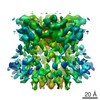
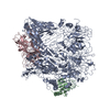
- Structure visualization
| Movie |
Movie viewer |
|---|---|
| Structure viewer | Molecule: MolmilJmol/JSmol |
 UCSF Chimera
- Downloads & links
-Download
| PDBx/mmCIF format | 6yeg.cif.gz | 343.5 KB | Display | PDBx/mmCIF format |
|---|---|---|---|---|
| PDB format | pdb6yeg.ent.gz | 283.6 KB | Display | PDB format |
| PDBx/mmJSON format | 6yeg.json.gz | Tree view | PDBx/mmJSON format | |
| Others |  Other downloads Other downloads |
-Validation report
| Arichive directory | https://data.pdbj.org/pub/pdb/validation_reports/ye/6yegftp://data.pdbj.org/pub/pdb/validation_reports/ye/6yeg | HTTPS FTP |
|---|
-Related structure data
| Related structure data |  10792MC 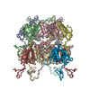 6yq5C M: map data used to model this data C: citing same article ( |
|---|---|
| Similar structure data | |
| Other databases |


-Links
 PDBj
PDBj


- Assembly
| Deposited unit | 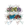
| |||||||||
|---|---|---|---|---|---|---|---|---|---|---|
| 1 |
| |||||||||
| NMR ensembles |
|
-Components
| #1: Protein | Mass: 19674.451 Da / Num. of mol.: 12 Source method: isolated from a genetically manipulated source Source: (gene. exp.) Bacillus phage SPP1 (virus) / Gene: 17.1 / Production host:  |
|---|
-Experimental details
-Experiment
| Experiment |
| ||||||||||||||||||||||||||||||||||||||||||||||||||||||||||||||||||||||||||||||||||||||||||||||||||||||||||||||||||
|---|---|---|---|---|---|---|---|---|---|---|---|---|---|---|---|---|---|---|---|---|---|---|---|---|---|---|---|---|---|---|---|---|---|---|---|---|---|---|---|---|---|---|---|---|---|---|---|---|---|---|---|---|---|---|---|---|---|---|---|---|---|---|---|---|---|---|---|---|---|---|---|---|---|---|---|---|---|---|---|---|---|---|---|---|---|---|---|---|---|---|---|---|---|---|---|---|---|---|---|---|---|---|---|---|---|---|---|---|---|---|---|---|---|---|---|
| EM experiment | Aggregation state: HELICAL ARRAY / 3D reconstruction method: helical reconstruction | ||||||||||||||||||||||||||||||||||||||||||||||||||||||||||||||||||||||||||||||||||||||||||||||||||||||||||||||||||
| NMR experiment |
|
- Sample preparation
| Component | Name: SPP1 tail tube / Type: COMPLEX / Entity ID: all / Source: RECOMBINANT | |||||||||||||||||||||||||||||||||||||||||||||||||||||||||||||||||||||||||||
|---|---|---|---|---|---|---|---|---|---|---|---|---|---|---|---|---|---|---|---|---|---|---|---|---|---|---|---|---|---|---|---|---|---|---|---|---|---|---|---|---|---|---|---|---|---|---|---|---|---|---|---|---|---|---|---|---|---|---|---|---|---|---|---|---|---|---|---|---|---|---|---|---|---|---|---|---|
| Source (natural) | Organism: Bacillus phage SPP1 (virus) | |||||||||||||||||||||||||||||||||||||||||||||||||||||||||||||||||||||||||||
| Source (recombinant) | Organism: | |||||||||||||||||||||||||||||||||||||||||||||||||||||||||||||||||||||||||||
| Buffer solution | pH: 7.4 | |||||||||||||||||||||||||||||||||||||||||||||||||||||||||||||||||||||||||||
| Buffer component |
| |||||||||||||||||||||||||||||||||||||||||||||||||||||||||||||||||||||||||||
| Specimen | Embedding applied: NO / Shadowing applied: NO / Staining applied: NO / Vitrification applied: YES | |||||||||||||||||||||||||||||||||||||||||||||||||||||||||||||||||||||||||||
| Vitrification | Cryogen name: ETHANE | |||||||||||||||||||||||||||||||||||||||||||||||||||||||||||||||||||||||||||
| Details |
| |||||||||||||||||||||||||||||||||||||||||||||||||||||||||||||||||||||||||||
| Sample |
|