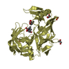
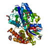
 Movie
Movie Controller


+ Open data

- Basic information
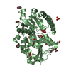
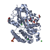
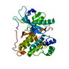
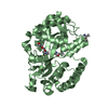
| Entry | Database: PDB / ID: 6ye8 | ||||||
|---|---|---|---|---|---|---|---|
| Title | E.coli's Putrescine receptor PotF complexed with Spermidine | ||||||
 Components Components | Putrescine-binding periplasmic protein | ||||||
 Keywords Keywords | TRANSPORT PROTEIN / Periplasmic binding protein / E.coli / Spermidine | ||||||
| Function / homology |  Function and homology information Function and homology informationspermidine binding / putrescine binding / putrescine transport / outer membrane-bounded periplasmic space / membrane Similarity search - Function | ||||||
| Biological species |  | ||||||
| Method |  X-RAY DIFFRACTION / SYNCHROTRON / MOLECULAR REPLACEMENT / Resolution: 1.5 Å X-RAY DIFFRACTION / SYNCHROTRON / MOLECULAR REPLACEMENT / Resolution: 1.5 Å | ||||||
 Authors Authors | Shanmugaratnam, S. / Kroeger, P. / Hocker, B. | ||||||
| Funding support |  Germany, 1items Germany, 1items
| ||||||
 Citation Citation | Journal: Structure / Year: 2021 Title: A comprehensive binding study illustrates ligand recognition in the periplasmic binding protein PotF. Authors: Kroger, P. / Shanmugaratnam, S. / Ferruz, N. / Schweimer, K. / Hocker, B. | ||||||
| History |
|
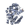
- Structure visualization
| Structure viewer | Molecule: MolmilJmol/JSmol |
|---|
- Downloads & links
-Download
| PDBx/mmCIF format | 6ye8.cif.gz | 509.9 KB | Display | PDBx/mmCIF format |
|---|---|---|---|---|
| PDB format | pdb6ye8.ent.gz | 351.1 KB | Display | PDB format |
| PDBx/mmJSON format | 6ye8.json.gz | Tree view | PDBx/mmJSON format | |
| Others |  Other downloads Other downloads |
-Validation report
| Arichive directory | https://data.pdbj.org/pub/pdb/validation_reports/ye/6ye8ftp://data.pdbj.org/pub/pdb/validation_reports/ye/6ye8 | HTTPS FTP |
|---|
-Related structure data
| Related structure data |  6ye0C 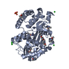 6ye6C  6ye7C  6yebC 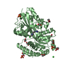 6yecC  6yedC 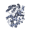 1a99S C: citing same article ( S: Starting model for refinement |
|---|---|
| Similar structure data |


-Links
 PDBj
PDBj


- Assembly
| Deposited unit | 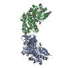
| ||||||||||||
|---|---|---|---|---|---|---|---|---|---|---|---|---|---|
| 1 | 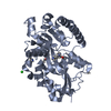
| ||||||||||||
| 2 | 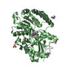
| ||||||||||||
| Unit cell |
|
-Components
-Protein , 1 types, 2 molecules AB
| #1: Protein | Mass: 39370.531 Da / Num. of mol.: 2 Source method: isolated from a genetically manipulated source Source: (gene. exp.) |
|---|
-Non-polymers , 6 types, 613 molecules 
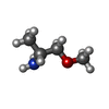
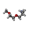


| #2: Chemical |  Mass: 145.246 Da / Num. of mol.: 2 / Source method: obtained synthetically / Formula: C7H19N3 / Feature type: SUBJECT OF INVESTIGATION Mass: 145.246 Da / Num. of mol.: 2 / Source method: obtained synthetically / Formula: C7H19N3 / Feature type: SUBJECT OF INVESTIGATION#3: Chemical | ChemComp-JFN / (  Mass: 89.136 Da / Num. of mol.: 7 / Source method: obtained synthetically / Formula: C4H11NO Mass: 89.136 Da / Num. of mol.: 7 / Source method: obtained synthetically / Formula: C4H11NO#4: Chemical |  Mass: 133.189 Da / Num. of mol.: 2 / Source method: obtained synthetically / Formula: C6H15NO2 Mass: 133.189 Da / Num. of mol.: 2 / Source method: obtained synthetically / Formula: C6H15NO2#5: Chemical |  Mass: 35.453 Da / Num. of mol.: 2 / Source method: obtained synthetically / Formula: Cl Mass: 35.453 Da / Num. of mol.: 2 / Source method: obtained synthetically / Formula: Cl#6: Chemical | ChemComp-SO4 / |  Mass: 96.063 Da / Num. of mol.: 1 / Source method: obtained synthetically / Formula: SO4 Mass: 96.063 Da / Num. of mol.: 1 / Source method: obtained synthetically / Formula: SO4#7: Water | ChemComp-HOH / | Mass: 18.015 Da / Num. of mol.: 599 / Source method: isolated from a natural source / Formula: H2O |
|---|
-Details
| Has ligand of interest | Y |
|---|---|
| Has protein modification | Y |
-Experimental details
-Experiment
| Experiment | Method: X-RAY DIFFRACTION / Number of used crystals: 1 |
|---|
- Sample preparation
| Crystal | Density Matthews: 2.5 Å3/Da / Density % sol: 50.84 % |
|---|---|
| Crystal grow | Temperature: 293 K / Method: vapor diffusion, sitting drop / pH: 8.8 Details: 2.4 M Ammoniumsulfate, 0.1 M Bicine pH 8.8, 10% Jeffamine M600 |
-Data collection
| Diffraction | Mean temperature: 100 K / Serial crystal experiment: N |
|---|---|
| Diffraction source | Source: SYNCHROTRON / Site: SLS  / Beamline: X06DA / Wavelength: 1 Å / Beamline: X06DA / Wavelength: 1 Å |
| Detector | Type: DECTRIS PILATUS 2M-F / Detector: PIXEL / Date: Oct 13, 2018 Details: vertically collimating mirror (M1, focus at infinity) |
| Radiation | Monochromator: Bartels Monochromator with dual channel cut crystals (DCCM) in (+--+) geometry and a toroidal mirror (M2) Protocol: SINGLE WAVELENGTH / Monochromatic (M) / Laue (L): M / Scattering type: x-ray |
| Radiation wavelength | Wavelength: 1 Å / Relative weight: 1 |
| Reflection | Resolution: 1.5→40.71 Å / Num. obs: 128236 / % possible obs: 99.97 % / Redundancy: 11.5 % / Biso Wilson estimate: 25.35 Å2 / CC1/2: 1 / CC star: 1 / Rmerge(I) obs: 0.07125 / Rpim(I) all: 0.02191 / Rrim(I) all: 0.07461 / Net I/σ(I): 17.79 |
| Reflection shell | Resolution: 1.5→1.554 Å / Redundancy: 11 % / Rmerge(I) obs: 2.663 / Mean I/σ(I) obs: 0.76 / Num. unique obs: 12611 / CC1/2: 0.264 / CC star: 0.647 / Rpim(I) all: 0.8379 / Rrim(I) all: 2.794 / % possible all: 99.99 |
- Processing
| Software |
| ||||||||||||||||||||||||||||||||||||||||||||||||||||||||||||||||||||||||||||||||||||||||||||||||||||||||||||||||
|---|---|---|---|---|---|---|---|---|---|---|---|---|---|---|---|---|---|---|---|---|---|---|---|---|---|---|---|---|---|---|---|---|---|---|---|---|---|---|---|---|---|---|---|---|---|---|---|---|---|---|---|---|---|---|---|---|---|---|---|---|---|---|---|---|---|---|---|---|---|---|---|---|---|---|---|---|---|---|---|---|---|---|---|---|---|---|---|---|---|---|---|---|---|---|---|---|---|---|---|---|---|---|---|---|---|---|---|---|---|---|---|---|---|
| Refinement | Method to determine structure: MOLECULAR REPLACEMENT Starting model: 1A99 Resolution: 1.5→40.71 Å / SU ML: 0.1903 / Cross valid method: FREE R-VALUE / σ(F): 1.33 / Phase error: 19.9395
| ||||||||||||||||||||||||||||||||||||||||||||||||||||||||||||||||||||||||||||||||||||||||||||||||||||||||||||||||
| Solvent computation | Shrinkage radii: 0.9 Å / VDW probe radii: 1.11 Å | ||||||||||||||||||||||||||||||||||||||||||||||||||||||||||||||||||||||||||||||||||||||||||||||||||||||||||||||||
| Displacement parameters | Biso mean: 33.69 Å2 | ||||||||||||||||||||||||||||||||||||||||||||||||||||||||||||||||||||||||||||||||||||||||||||||||||||||||||||||||
| Refinement step | Cycle: LAST / Resolution: 1.5→40.71 Å
| ||||||||||||||||||||||||||||||||||||||||||||||||||||||||||||||||||||||||||||||||||||||||||||||||||||||||||||||||
| Refine LS restraints |
| ||||||||||||||||||||||||||||||||||||||||||||||||||||||||||||||||||||||||||||||||||||||||||||||||||||||||||||||||
| LS refinement shell |
| ||||||||||||||||||||||||||||||||||||||||||||||||||||||||||||||||||||||||||||||||||||||||||||||||||||||||||||||||
| Refinement TLS params. | Method: refined / Refine-ID: X-RAY DIFFRACTION
| ||||||||||||||||||||||||||||||||||||||||||||||||||||||||||||||||||||||||||||||||||||||||||||||||||||||||||||||||
| Refinement TLS group |
|
