 Movie
Movie Controller

[English] 日本語
 Yorodumi
- PDB-6yby: Crystal structure of the D116N mutant of the light-driven sodium ... -
+ Open data

- Basic information
| Entry | Database: PDB / ID: 6yby | ||||||||||||||||||
|---|---|---|---|---|---|---|---|---|---|---|---|---|---|---|---|---|---|---|---|
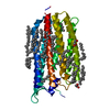
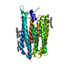
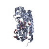
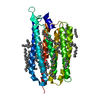
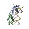
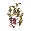
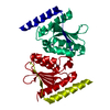
| Title | Crystal structure of the D116N mutant of the light-driven sodium pump KR2 in the monomeric form, pH 4.6 | ||||||||||||||||||
 Components Components | Sodium pumping rhodopsin | ||||||||||||||||||
 Keywords Keywords | MEMBRANE PROTEIN / rhodopsin / ion pumping / retinal / sodium pump / intermediate state | ||||||||||||||||||
| Function / homology |  Function and homology information Function and homology information | ||||||||||||||||||
| Biological species |  Dokdonia eikasta (bacteria) Dokdonia eikasta (bacteria) | ||||||||||||||||||
| Method |  X-RAY DIFFRACTION / SYNCHROTRON / MOLECULAR REPLACEMENT / Resolution: 1.8 Å X-RAY DIFFRACTION / SYNCHROTRON / MOLECULAR REPLACEMENT / Resolution: 1.8 Å | ||||||||||||||||||
 Authors Authors | Kovalev, K. / Gushchin, I. / Gordeliy, V. | ||||||||||||||||||
| Funding support |  France, France,  Russian Federation, 5items Russian Federation, 5items
| ||||||||||||||||||
 Citation Citation | Journal: Nat Commun / Year: 2020 Title: Molecular mechanism of light-driven sodium pumping. Authors: Kovalev, K. / Astashkin, R. / Gushchin, I. / Orekhov, P. / Volkov, D. / Zinovev, E. / Marin, E. / Rulev, M. / Alekseev, A. / Royant, A. / Carpentier, P. / Vaganova, S. / Zabelskii, D. / ...Authors: Kovalev, K. / Astashkin, R. / Gushchin, I. / Orekhov, P. / Volkov, D. / Zinovev, E. / Marin, E. / Rulev, M. / Alekseev, A. / Royant, A. / Carpentier, P. / Vaganova, S. / Zabelskii, D. / Baeken, C. / Sergeev, I. / Balandin, T. / Bourenkov, G. / Carpena, X. / Boer, R. / Maliar, N. / Borshchevskiy, V. / Buldt, G. / Bamberg, E. / Gordeliy, V. | ||||||||||||||||||
| History |
|
- Structure visualization
| Structure viewer | Molecule: MolmilJmol/JSmol |
|---|
- Downloads & links
-Download
| PDBx/mmCIF format | 6yby.cif.gz | 87.1 KB | Display | PDBx/mmCIF format |
|---|---|---|---|---|
| PDB format | pdb6yby.ent.gz | 62.2 KB | Display | PDB format |
| PDBx/mmJSON format | 6yby.json.gz | Tree view | PDBx/mmJSON format | |
| Others |  Other downloads Other downloads |
-Validation report
| Arichive directory | https://data.pdbj.org/pub/pdb/validation_reports/yb/6ybyftp://data.pdbj.org/pub/pdb/validation_reports/yb/6yby | HTTPS FTP |
|---|
-Related structure data
| Related structure data |  6xytC  6ybzC  6yc0C  6yc1C  6yc2C  6yc3C  6yc4C  4xtlS S: Starting model for refinement C: citing same article ( |
|---|---|
| Similar structure data |
-Links
 PDBj
PDBj


- Assembly
| Deposited unit | 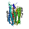
| ||||||||||||
|---|---|---|---|---|---|---|---|---|---|---|---|---|---|
| 1 |
| ||||||||||||
| Unit cell |
| ||||||||||||
| Components on special symmetry positions |
|
-Components
| #1: Protein | Mass: 30907.756 Da / Num. of mol.: 1 Source method: isolated from a genetically manipulated source Source: (gene. exp.) Dokdonia eikasta (bacteria) / Gene: NaR / Production host: | ||||||||
|---|---|---|---|---|---|---|---|---|---|
| #2: Chemical | ChemComp-NA /   Mass: 22.990 Da / Num. of mol.: 1 / Source method: obtained synthetically / Formula: Na / Feature type: SUBJECT OF INVESTIGATION Mass: 22.990 Da / Num. of mol.: 1 / Source method: obtained synthetically / Formula: Na / Feature type: SUBJECT OF INVESTIGATION | ||||||||
| #3: Chemical | ChemComp-LFA /   Mass: 282.547 Da / Num. of mol.: 34 / Source method: obtained synthetically / Formula: C20H42 Mass: 282.547 Da / Num. of mol.: 34 / Source method: obtained synthetically / Formula: C20H42#4: Chemical | ChemComp-RET / |   Mass: 284.436 Da / Num. of mol.: 1 / Source method: obtained synthetically / Formula: C20H28O Mass: 284.436 Da / Num. of mol.: 1 / Source method: obtained synthetically / Formula: C20H28O#5: Water | ChemComp-HOH / |  Mass: 18.015 Da / Num. of mol.: 151 / Source method: isolated from a natural source / Formula: H2O Mass: 18.015 Da / Num. of mol.: 151 / Source method: isolated from a natural source / Formula: H2OHas ligand of interest | Y | Has protein modification | Y | |
-Experimental details
-Experiment
| Experiment | Method: X-RAY DIFFRACTION / Number of used crystals: 1 |
|---|
- Sample preparation
| Crystal | Density Matthews: 3.23 Å3/Da / Density % sol: 61.95 % |
|---|---|
| Crystal grow | Temperature: 293 K / Method: lipidic cubic phase / pH: 4.6 / Details: 2 M NaMal pH 4.6 |
-Data collection
| Diffraction | Mean temperature: 100 K / Serial crystal experiment: N | ||||||||||||||||||||||||||||||
|---|---|---|---|---|---|---|---|---|---|---|---|---|---|---|---|---|---|---|---|---|---|---|---|---|---|---|---|---|---|---|---|
| Diffraction source | Source: SYNCHROTRON / Site: ESRF / Beamline: ID30B / Wavelength: 0.978 Å | ||||||||||||||||||||||||||||||
| Detector | Type: DECTRIS PILATUS3 S 6M / Detector: PIXEL / Date: Aug 26, 2018 | ||||||||||||||||||||||||||||||
| Radiation | Protocol: SINGLE WAVELENGTH / Monochromatic (M) / Laue (L): M / Scattering type: x-ray | ||||||||||||||||||||||||||||||
| Radiation wavelength | Wavelength: 0.978 Å / Relative weight: 1 | ||||||||||||||||||||||||||||||
| Reflection | Resolution: 1.8→40.81 Å / Num. obs: 37771 / % possible obs: 99.8 % / Redundancy: 6.6 % / CC1/2: 0.999 / Rmerge(I) obs: 0.046 / Rpim(I) all: 0.02 / Rrim(I) all: 0.05 / Net I/σ(I): 16.2 / Num. measured all: 249134 / Scaling rejects: 12 | ||||||||||||||||||||||||||||||
| Reflection shell | Diffraction-ID: 1
|
- Processing
| Software |
| ||||||||||||||||||||||||||||||||||||||||||||||||||||||||||||
|---|---|---|---|---|---|---|---|---|---|---|---|---|---|---|---|---|---|---|---|---|---|---|---|---|---|---|---|---|---|---|---|---|---|---|---|---|---|---|---|---|---|---|---|---|---|---|---|---|---|---|---|---|---|---|---|---|---|---|---|---|---|
| Refinement | Method to determine structure: MOLECULAR REPLACEMENT Starting model: 4xtl Resolution: 1.8→19.83 Å / Cor.coef. Fo:Fc: 0.961 / Cor.coef. Fo:Fc free: 0.961 / SU B: 3.571 / SU ML: 0.1 / Cross valid method: THROUGHOUT / σ(F): 0 / ESU R: 0.121 / ESU R Free: 0.111 Details: HYDROGENS HAVE BEEN ADDED IN THE RIDING POSITIONS U VALUES : REFINED INDIVIDUALLY
| ||||||||||||||||||||||||||||||||||||||||||||||||||||||||||||
| Solvent computation | Ion probe radii: 0.8 Å / Shrinkage radii: 0.8 Å / VDW probe radii: 1.2 Å | ||||||||||||||||||||||||||||||||||||||||||||||||||||||||||||
| Displacement parameters | Biso max: 170.56 Å2 / Biso mean: 43.739 Å2 / Biso min: 21.65 Å2
| ||||||||||||||||||||||||||||||||||||||||||||||||||||||||||||
| Refinement step | Cycle: final / Resolution: 1.8→19.83 Å
| ||||||||||||||||||||||||||||||||||||||||||||||||||||||||||||
| Refine LS restraints |
| ||||||||||||||||||||||||||||||||||||||||||||||||||||||||||||
| LS refinement shell | Resolution: 1.8→1.846 Å / Rfactor Rfree error: 0 / Total num. of bins used: 20
|
