 Movie
Movie Controller

[English] 日本語
 Yorodumi
- PDB-6yc2: Crystal structure of the light-driven sodium pump KR2 in the pent... -
+ Open data

- Basic information
| Entry | Database: PDB / ID: 6yc2 | ||||||||||||||||||
|---|---|---|---|---|---|---|---|---|---|---|---|---|---|---|---|---|---|---|---|
| Title | Crystal structure of the light-driven sodium pump KR2 in the pentameric form at room temperature, pH 8.0 | ||||||||||||||||||
 Components Components | Sodium pumping rhodopsin | ||||||||||||||||||
 Keywords Keywords | MEMBRANE PROTEIN / rhodopsin / ion pumping / retinal / sodium pump / intermediate state | ||||||||||||||||||
| Function / homology | Bacteriorhodopsin-like protein / Archaeal/bacterial/fungal rhodopsins / Bacteriorhodopsin-like protein / membrane / ALANINE / EICOSANE / OLEIC ACID / (2R)-2,3-dihydroxypropyl (9Z)-octadec-9-enoate / Sodium pumping rhodopsin Function and homology information Function and homology information | ||||||||||||||||||
| Biological species |  Dokdonia eikasta (bacteria) Dokdonia eikasta (bacteria) | ||||||||||||||||||
| Method |  X-RAY DIFFRACTION / SYNCHROTRON / MOLECULAR REPLACEMENT / Resolution: 2.5 Å X-RAY DIFFRACTION / SYNCHROTRON / MOLECULAR REPLACEMENT / Resolution: 2.5 Å | ||||||||||||||||||
 Authors Authors | Kovalev, K. / Gushchin, I. / Gordeliy, V. | ||||||||||||||||||
| Funding support |  France, France,  Russian Federation, 5items Russian Federation, 5items
| ||||||||||||||||||
 Citation Citation | Journal: Nat Commun / Year: 2020 Title: Molecular mechanism of light-driven sodium pumping. Authors: Kovalev, K. / Astashkin, R. / Gushchin, I. / Orekhov, P. / Volkov, D. / Zinovev, E. / Marin, E. / Rulev, M. / Alekseev, A. / Royant, A. / Carpentier, P. / Vaganova, S. / Zabelskii, D. / ...Authors: Kovalev, K. / Astashkin, R. / Gushchin, I. / Orekhov, P. / Volkov, D. / Zinovev, E. / Marin, E. / Rulev, M. / Alekseev, A. / Royant, A. / Carpentier, P. / Vaganova, S. / Zabelskii, D. / Baeken, C. / Sergeev, I. / Balandin, T. / Bourenkov, G. / Carpena, X. / Boer, R. / Maliar, N. / Borshchevskiy, V. / Buldt, G. / Bamberg, E. / Gordeliy, V. | ||||||||||||||||||
| History |
|
- Structure visualization
| Structure viewer | Molecule: MolmilJmol/JSmol |
|---|
- Downloads & links
-Download
| PDBx/mmCIF format | 6yc2.cif.gz | 291.2 KB | Display | PDBx/mmCIF format |
|---|---|---|---|---|
| PDB format | pdb6yc2.ent.gz | 235.1 KB | Display | PDB format |
| PDBx/mmJSON format | 6yc2.json.gz | Tree view | PDBx/mmJSON format | |
| Others |  Other downloads Other downloads |
-Validation report
| Arichive directory | https://data.pdbj.org/pub/pdb/validation_reports/yc/6yc2ftp://data.pdbj.org/pub/pdb/validation_reports/yc/6yc2 | HTTPS FTP |
|---|
-Related structure data
| Related structure data |  6xytC 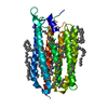 6ybyC  6ybzC  6yc0C  6yc1C  6yc3C  6yc4C  6rew S: Starting model for refinement C: citing same article ( |
|---|---|
| Similar structure data |


-Links
 PDBj
PDBj


- Assembly
| Deposited unit | 
| |||||||||||||||||||||||||||||||||||||||||||||||||||||||||||||||||||||||||||||||||||||||||||||||||||||||||||||||||||||||||||||||||||||||||||||||||||||||||||||||||||||||||||||||||||||||||||||||||||||||||||||||||||||||||||||
|---|---|---|---|---|---|---|---|---|---|---|---|---|---|---|---|---|---|---|---|---|---|---|---|---|---|---|---|---|---|---|---|---|---|---|---|---|---|---|---|---|---|---|---|---|---|---|---|---|---|---|---|---|---|---|---|---|---|---|---|---|---|---|---|---|---|---|---|---|---|---|---|---|---|---|---|---|---|---|---|---|---|---|---|---|---|---|---|---|---|---|---|---|---|---|---|---|---|---|---|---|---|---|---|---|---|---|---|---|---|---|---|---|---|---|---|---|---|---|---|---|---|---|---|---|---|---|---|---|---|---|---|---|---|---|---|---|---|---|---|---|---|---|---|---|---|---|---|---|---|---|---|---|---|---|---|---|---|---|---|---|---|---|---|---|---|---|---|---|---|---|---|---|---|---|---|---|---|---|---|---|---|---|---|---|---|---|---|---|---|---|---|---|---|---|---|---|---|---|---|---|---|---|---|---|---|---|---|---|---|---|---|---|---|---|---|---|---|---|---|---|---|---|
| 1 |
| |||||||||||||||||||||||||||||||||||||||||||||||||||||||||||||||||||||||||||||||||||||||||||||||||||||||||||||||||||||||||||||||||||||||||||||||||||||||||||||||||||||||||||||||||||||||||||||||||||||||||||||||||||||||||||||
| Unit cell |
| |||||||||||||||||||||||||||||||||||||||||||||||||||||||||||||||||||||||||||||||||||||||||||||||||||||||||||||||||||||||||||||||||||||||||||||||||||||||||||||||||||||||||||||||||||||||||||||||||||||||||||||||||||||||||||||
| Noncrystallographic symmetry (NCS) | NCS domain:
NCS domain segments: Component-ID: _ / Beg auth comp-ID: GLN / Beg label comp-ID: GLN / Refine code: _
|