 Movie
Movie Controller

[English] 日本語
 Yorodumi
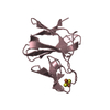
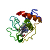
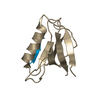
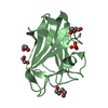
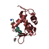
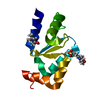
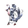
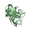
- PDB-6y9n: Crystal structure of Whirlin PDZ3_C-ter in complex with Myosin 15... -
+ Open data

- Basic information
| Entry | Database: PDB / ID: 6y9n | ||||||
|---|---|---|---|---|---|---|---|
| Title | Crystal structure of Whirlin PDZ3_C-ter in complex with Myosin 15a C-terminal PDZ binding motif peptide | ||||||
 Components Components |
| ||||||
 Keywords Keywords | STRUCTURAL PROTEIN / Whirlin / PDZ / Myosin 15a / complex | ||||||
| Function / homology |  Function and homology information Function and homology informationaxon collateral / paranodal junction maintenance / actin-based cell projection / USH2 complex / stereocilia ankle link / periciliary membrane compartment / stereocilia ankle link complex / inner ear receptor cell differentiation / cerebellar Purkinje cell layer formation / sensory perception of light stimulus ...axon collateral / paranodal junction maintenance / actin-based cell projection / USH2 complex / stereocilia ankle link / periciliary membrane compartment / stereocilia ankle link complex / inner ear receptor cell differentiation / cerebellar Purkinje cell layer formation / sensory perception of light stimulus / inner ear receptor cell stereocilium organization / stereocilium tip / photoreceptor connecting cilium / stereocilium bundle / detection of mechanical stimulus involved in sensory perception of sound / stereocilium / retina homeostasis / apical dendrite / auditory receptor cell stereocilium organization / myosin complex / inner ear morphogenesis / microfilament motor activity / response to light stimulus / photoreceptor inner segment / actin filament organization / dendritic shaft / actin filament / establishment of protein localization / locomotory behavior / sensory perception of sound / endocytosis / actin filament binding / actin cytoskeleton / growth cone / presynapse / postsynapse / cilium / ciliary basal body / protein domain specific binding / neuronal cell body / positive regulation of gene expression / perinuclear region of cytoplasm / ATP binding / membrane / identical protein binding / plasma membrane / cytoplasm / cytosol Similarity search - Function | ||||||
| Biological species |  | ||||||
| Method |  X-RAY DIFFRACTION / SYNCHROTRON / MOLECULAR REPLACEMENT / Resolution: 1.93 Å X-RAY DIFFRACTION / SYNCHROTRON / MOLECULAR REPLACEMENT / Resolution: 1.93 Å | ||||||
 Authors Authors | Zhu, Y. / Delhommel, F. / Haouz, A. / Caillet-Saguy, C. / Vaney, M. / Mechaly, A.E. / Wolff, N. | ||||||
| Funding support |  France, 1items France, 1items
| ||||||
 Citation Citation | Journal: J.Mol.Biol. / Year: 2020 Title: Deciphering the Unexpected Binding Capacity of the Third PDZ Domain of Whirlin to Various Cochlear Hair Cell Partners. Authors: Zhu, Y. / Delhommel, F. / Cordier, F. / Luchow, S. / Mechaly, A. / Colcombet-Cazenave, B. / Girault, V. / Pepermans, E. / Bahloul, A. / Gautier, C. / Brule, S. / Raynal, B. / Hoos, S. / ...Authors: Zhu, Y. / Delhommel, F. / Cordier, F. / Luchow, S. / Mechaly, A. / Colcombet-Cazenave, B. / Girault, V. / Pepermans, E. / Bahloul, A. / Gautier, C. / Brule, S. / Raynal, B. / Hoos, S. / Haouz, A. / Caillet-Saguy, C. / Ivarsson, Y. / Wolff, N. | ||||||
| History |
|
- Structure visualization
| Structure viewer | Molecule: MolmilJmol/JSmol |
|---|
- Downloads & links
-Download
| PDBx/mmCIF format | 6y9n.cif.gz | 59.5 KB | Display | PDBx/mmCIF format |
|---|---|---|---|---|
| PDB format | pdb6y9n.ent.gz | 40.7 KB | Display | PDB format |
| PDBx/mmJSON format | 6y9n.json.gz | Tree view | PDBx/mmJSON format | |
| Others |  Other downloads Other downloads |
-Validation report
| Arichive directory | https://data.pdbj.org/pub/pdb/validation_reports/y9/6y9nftp://data.pdbj.org/pub/pdb/validation_reports/y9/6y9n | HTTPS FTP |
|---|
-Related structure data
| Related structure data |  6y38C  6y9oC 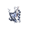 6y9pC 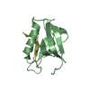 6y9qC 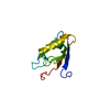 1ufxS S: Starting model for refinement C: citing same article ( |
|---|---|
| Similar structure data |


-Links
 PDBj
PDBj


- Assembly
| Deposited unit | 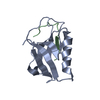
| ||||||||||||
|---|---|---|---|---|---|---|---|---|---|---|---|---|---|
| 1 |
| ||||||||||||
| Unit cell |
|
-Components
| #1: Protein | Mass: 11359.070 Da / Num. of mol.: 1 Source method: isolated from a genetically manipulated source Source: (gene. exp.) Production host:  References: UniProt: Q80VW5 |
|---|---|
| #2: Protein/peptide | Mass: 1482.741 Da / Num. of mol.: 1 / Source method: obtained synthetically / Source: (synth.) |
| #3: Water | ChemComp-HOH /  Mass: 18.015 Da / Num. of mol.: 75 / Source method: isolated from a natural source / Formula: H2O Mass: 18.015 Da / Num. of mol.: 75 / Source method: isolated from a natural source / Formula: H2O |
-Experimental details
-Experiment
| Experiment | Method: X-RAY DIFFRACTION / Number of used crystals: 1 |
|---|
- Sample preparation
| Crystal | Density Matthews: 3.8 Å3/Da / Density % sol: 67.6 % |
|---|---|
| Crystal grow | Temperature: 277.15 K / Method: vapor diffusion, sitting drop Details: 0.1M HEPES pH 7.5,10% v/v isopropanol,20% w/v PEG 4000 |
-Data collection
| Diffraction | Mean temperature: 100 K / Serial crystal experiment: N |
|---|---|
| Diffraction source | Source: SYNCHROTRON / Site: SOLEIL / Beamline: PROXIMA 1 / Wavelength: 0.9786 Å |
| Detector | Type: DECTRIS PILATUS 300K / Detector: PIXEL / Date: Sep 29, 2017 |
| Radiation | Protocol: SINGLE WAVELENGTH / Monochromatic (M) / Laue (L): M / Scattering type: x-ray |
| Radiation wavelength | Wavelength: 0.9786 Å / Relative weight: 1 |
| Reflection | Resolution: 1.93→15.7 Å / Num. obs: 14149 / % possible obs: 99.38 % / Redundancy: 7.2 % / CC1/2: 0.998 / CC star: 1 / Net I/σ(I): 13.01 |
| Reflection shell | Resolution: 1.93→1.999 Å / Num. unique obs: 1403 / CC1/2: 0.659 / CC star: 0.891 |
- Processing
| Software |
| ||||||||||||||||||||||||
|---|---|---|---|---|---|---|---|---|---|---|---|---|---|---|---|---|---|---|---|---|---|---|---|---|---|
| Refinement | Method to determine structure: MOLECULAR REPLACEMENT Starting model: 1UFX Resolution: 1.93→15.7 Å / Cross valid method: FREE R-VALUE
| ||||||||||||||||||||||||
| Displacement parameters | Biso mean: 48.37 Å2 | ||||||||||||||||||||||||
| Refinement step | Cycle: LAST / Resolution: 1.93→15.7 Å
| ||||||||||||||||||||||||
| Refine LS restraints |
|
