Movie
Movie Controller

[English] 日本語
 Yorodumi
- PDB-6xzc: CryoEM structure of the ring-shaped virulence factor EspB from My... -
+ Open data

- Basic information
| Entry | Database: PDB / ID: 6xzc | ||||||
|---|---|---|---|---|---|---|---|
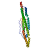
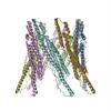
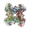
| Title | CryoEM structure of the ring-shaped virulence factor EspB from Mycobacterium tuberculosis | ||||||
 Components Components | ESX-1 secretion-associated protein EspB | ||||||
 Keywords Keywords | TRANSPORT PROTEIN / M. tuberculosis / ESX-1 / Type VII secretion system / EspB / Rv3881c / EsxA / CFP10 / ESAT-6 | ||||||
| Function / homology |  Function and homology information Function and homology informationprotein secretion by the type VII secretion system / biological process involved in interaction with host / extracellular region / identical protein binding Similarity search - Function | ||||||
| Biological species |   Mycobacterium tuberculosis (bacteria) Mycobacterium tuberculosis (bacteria) | ||||||
| Method | ELECTRON MICROSCOPY / single particle reconstruction / cryo EM / Resolution: 3.37 Å | ||||||
 Authors Authors | Piton, J. / Pojer, F. / Wakatsuki, S. / Gati, C. / Cole, S.T. | ||||||
| Funding support |  Switzerland, 1items Switzerland, 1items
| ||||||
 Citation Citation | Journal: J Struct Biol X / Year: 2020 Title: High resolution CryoEM structure of the ring-shaped virulence factor EspB from . Authors: Jérémie Piton / Florence Pojer / Soichi Wakatsuki / Cornelius Gati / Stewart T Cole /  Abstract: The EspB protein of is a 60 kDa virulence factor, implicated in conjugation and exported by the ESX-1 system of which it may also be a component. Previous attempts to obtain high-resolution maps of ...The EspB protein of is a 60 kDa virulence factor, implicated in conjugation and exported by the ESX-1 system of which it may also be a component. Previous attempts to obtain high-resolution maps of EspB by cryo-electron microscopic examination of single particles have been thwarted by severe orientation bias of the particles. This was overcome by using detergent as a surfactant thereby allowing reconstruction of the EspB structure at 3.37 Å resolution. The final structure revealed the N-terminal domain of EspB to be organized as a cylindrical heptamer with dimensions of 90 Å x 90 Å and a central channel of 45 Å diameter whereas the C-terminal domain was unstructured. New atomic insight was obtained into the helical packing required for protomer interactions and the overall electrostatic potential. The external surface is electronegatively charged while the channel is lined with electropositive patches. EspB thus has many features of a pore-like transport protein that might allow the passage of an ESX-1 substrate such as the 35 Å diameter EsxA-EsxB heterodimer or B-form DNA consistent with its proposed role in DNA uptake. | ||||||
| History |
|
- Structure visualization
| Movie |
Movie viewer |
|---|---|
| Structure viewer | Molecule: MolmilJmol/JSmol |
- Downloads & links
-Download
| PDBx/mmCIF format | 6xzc.cif.gz | 306.6 KB | Display | PDBx/mmCIF format |
|---|---|---|---|---|
| PDB format | pdb6xzc.ent.gz | 244.8 KB | Display | PDB format |
| PDBx/mmJSON format | 6xzc.json.gz | Tree view | PDBx/mmJSON format | |
| Others |  Other downloads Other downloads |
-Validation report
| Summary document | 6xzc_validation.pdf.gz | 1.4 MB | Display | wwPDB validaton report |
|---|---|---|---|---|
| Full document | 6xzc_full_validation.pdf.gz | 1.4 MB | Display | |
| Data in XML | 6xzc_validation.xml.gz | 54 KB | Display | |
| Data in CIF | 6xzc_validation.cif.gz | 74.1 KB | Display | |
| Arichive directory | https://data.pdbj.org/pub/pdb/validation_reports/xz/6xzcftp://data.pdbj.org/pub/pdb/validation_reports/xz/6xzc | HTTPS FTP |
-Related structure data
| Related structure data | 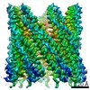 10658MC M: map data used to model this data C: citing same article ( |
|---|---|
| Similar structure data |


-Links
 PDBj
PDBj- Assembly
| Deposited unit | 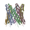
|
|---|---|
| 1 |
|
-Components
| #1: Protein | Mass: 47637.527 Da / Num. of mol.: 7 Source method: isolated from a genetically manipulated source Source: (gene. exp.) Mycobacterium tuberculosis (strain ATCC 25618 / H37Rv) (bacteria)Strain: ATCC 25618 / H37Rv / Gene: espB, mtb48, Rv3881c, MTV027.16c / Production host: |
|---|
-Experimental details
-Experiment
| Experiment | Method: ELECTRON MICROSCOPY |
|---|---|
| EM experiment | Aggregation state: PARTICLE / 3D reconstruction method: single particle reconstruction |
- Sample preparation
| Component | Name: EspB Heptameric complex / Type: COMPLEX / Entity ID: all / Source: RECOMBINANT | ||||||||||||
|---|---|---|---|---|---|---|---|---|---|---|---|---|---|
| Molecular weight | Value: 0.329 MDa / Experimental value: NO | ||||||||||||
| Source (natural) | Organism: Mycobacterium tuberculosis H37Rv (bacteria) | ||||||||||||
| Source (recombinant) | Organism: | ||||||||||||
| Buffer solution | pH: 8 | ||||||||||||
| Buffer component |
| ||||||||||||
| Specimen | Conc.: 5 mg/ml / Embedding applied: NO / Shadowing applied: NO / Staining applied: NO / Vitrification applied: YES / Details: This sample was monodiperse | ||||||||||||
| Specimen support | Grid material: GOLD / Grid mesh size: 300 divisions/in. / Grid type: Quantifoil R1.2/1.3 | ||||||||||||
| Vitrification | Instrument: FEI VITROBOT MARK IV / Cryogen name: ETHANE Details: 0.05% fluorinated octylmaltoside was add into protein solution and immediately applied to a freshly glow-discharged holey carbon grid. ). Excess liquid was blotted for 2.3 s using an FEI ...Details: 0.05% fluorinated octylmaltoside was add into protein solution and immediately applied to a freshly glow-discharged holey carbon grid. ). Excess liquid was blotted for 2.3 s using an FEI Vitrobot Mark IV and the sample was plunge frozen in liquid ethane. |
- Electron microscopy imaging
| Experimental equipment |  Model: Titan Krios / Image courtesy: FEI Company |
|---|---|
| Microscopy | Model: FEI TITAN KRIOS |
| Electron gun | Electron source:  FIELD EMISSION GUN / Accelerating voltage: 300 kV / Illumination mode: FLOOD BEAM FIELD EMISSION GUN / Accelerating voltage: 300 kV / Illumination mode: FLOOD BEAM |
| Electron lens | Mode: BRIGHT FIELD / Nominal defocus min: 1500 nm / Calibrated defocus max: 3000 nm / Cs: 2.7 mm |
| Image recording | Electron dose: 30 e/Å2 / Detector mode: SUPER-RESOLUTION / Film or detector model: GATAN K2 SUMMIT (4k x 4k) / Num. of real images: 2600 |
| EM imaging optics | Energyfilter name: GIF Quantum LS |
- Processing
| Software | Name: PHENIX / Version: 1.17.1_3660: / Classification: refinement | ||||||||||||||||||||||||||||||||||||||||
|---|---|---|---|---|---|---|---|---|---|---|---|---|---|---|---|---|---|---|---|---|---|---|---|---|---|---|---|---|---|---|---|---|---|---|---|---|---|---|---|---|---|
| EM software |
| ||||||||||||||||||||||||||||||||||||||||
| CTF correction | Type: PHASE FLIPPING AND AMPLITUDE CORRECTION | ||||||||||||||||||||||||||||||||||||||||
| Particle selection | Num. of particles selected: 292964 | ||||||||||||||||||||||||||||||||||||||||
| Symmetry | Point symmetry: C7 (7 fold cyclic) | ||||||||||||||||||||||||||||||||||||||||
| 3D reconstruction | Resolution: 3.37 Å / Resolution method: FSC 0.143 CUT-OFF / Num. of particles: 143350 / Num. of class averages: 50 / Symmetry type: POINT | ||||||||||||||||||||||||||||||||||||||||
| Atomic model building | Protocol: FLEXIBLE FIT / Space: REAL | ||||||||||||||||||||||||||||||||||||||||
| Atomic model building | PDB-ID: 4XXX Pdb chain-ID: A / Accession code: 4XXX / Source name: PDB / Type: experimental model | ||||||||||||||||||||||||||||||||||||||||
| Refine LS restraints |
|