 Movie
Movie Controller


+ Open data

- Basic information
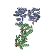
| Entry | Database: PDB / ID: 6xz4 | |||||||||
|---|---|---|---|---|---|---|---|---|---|---|
| Title | Crystal structure of TLNRD1 | |||||||||
 Components Components | Talin rod domain-containing protein 1 | |||||||||
 Keywords Keywords | CELL ADHESION / Actin binding protein / Protein complex / Cytoskeleton | |||||||||
| Function / homology | Talin rod domain-containing protein 1 / : / Talin IBS2B domain / stress fiber / actin binding / identical protein binding / Talin rod domain-containing protein 1 Function and homology information Function and homology information | |||||||||
| Biological species |  Homo sapiens (human) Homo sapiens (human) | |||||||||
| Method |  X-RAY DIFFRACTION / SYNCHROTRON / MOLECULAR REPLACEMENT / Resolution: 2.3 Å X-RAY DIFFRACTION / SYNCHROTRON / MOLECULAR REPLACEMENT / Resolution: 2.3 Å | |||||||||
 Authors Authors | Cowell, A. / Singh, A.K. / Brown, D.G. / Goult, B.T. | |||||||||
| Funding support |  United Kingdom, 2items United Kingdom, 2items
| |||||||||
 Citation Citation | Journal: J.Cell Biol. / Year: 2021 Title: Talin rod domain-containing protein 1 (TLNRD1) is a novel actin-bundling protein which promotes filopodia formation. Authors: Cowell, A.R. / Jacquemet, G. / Singh, A.K. / Varela, L. / Nylund, A.S. / Ammon, Y.C. / Brown, D.G. / Akhmanova, A. / Ivaska, J. / Goult, B.T. #1: Journal: Biorxiv / Year: 2020Title: Talin Rod Domain Containing Protein 1 (TLNRD1) is a novel actin- bundling protein which promotes filopodia formation. Authors: Cowell, A. / Jacquemet, G. / Singh, A.K. / Ammon, Y.-K. / Brown, D.G. / Akhmanova, A. / Ivaska, J. / Goult, B.T. | |||||||||
| History |
|
- Structure visualization
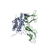
| Structure viewer | Molecule: MolmilJmol/JSmol |
|---|
- Downloads & links
-Download
| PDBx/mmCIF format | 6xz4.cif.gz | 222.6 KB | Display | PDBx/mmCIF format |
|---|---|---|---|---|
| PDB format | pdb6xz4.ent.gz | 180.9 KB | Display | PDB format |
| PDBx/mmJSON format | 6xz4.json.gz | Tree view | PDBx/mmJSON format | |
| Others |  Other downloads Other downloads |
-Validation report
| Arichive directory | https://data.pdbj.org/pub/pdb/validation_reports/xz/6xz4ftp://data.pdbj.org/pub/pdb/validation_reports/xz/6xz4 | HTTPS FTP |
|---|
-Related structure data
| Related structure data | 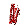 6xz3SC S: Starting model for refinement C: citing same article ( |
|---|---|
| Similar structure data |


-Links
 PDBj
PDBj- Assembly
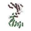
| Deposited unit | 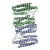
| |||||||||||||||||||||||||||||||||||||||
|---|---|---|---|---|---|---|---|---|---|---|---|---|---|---|---|---|---|---|---|---|---|---|---|---|---|---|---|---|---|---|---|---|---|---|---|---|---|---|---|---|
| 1 |
| |||||||||||||||||||||||||||||||||||||||
| Unit cell |
| |||||||||||||||||||||||||||||||||||||||
| Noncrystallographic symmetry (NCS) | NCS domain:
NCS domain segments: Component-ID: 1 / Ens-ID: 1
|
-Components
| #1: Protein | Mass: 38432.910 Da / Num. of mol.: 2 Source method: isolated from a genetically manipulated source Details: Full length TLNRD1 residues 1-362 / Source: (gene. exp.) Homo sapiens (human) / Gene: TLNRD1, MESDC1 / Plasmid: pET151 / Production host:  #2: Water | ChemComp-HOH / |  Mass: 18.015 Da / Num. of mol.: 70 / Source method: isolated from a natural source / Formula: H2O Mass: 18.015 Da / Num. of mol.: 70 / Source method: isolated from a natural source / Formula: H2O |
|---|
-Experimental details
-Experiment
| Experiment | Method: X-RAY DIFFRACTION / Number of used crystals: 1 |
|---|
- Sample preparation
| Crystal | Density Matthews: 2.12 Å3/Da / Density % sol: 41.98 % |
|---|---|
| Crystal grow | Temperature: 294 K / Method: vapor diffusion, hanging drop / Details: 250 mM sodium thiocyanate, 20% PEG 3350 |
-Data collection
| Diffraction | Mean temperature: 100 K / Serial crystal experiment: N |
|---|---|
| Diffraction source | Source: SYNCHROTRON / Site: SOLEIL  / Beamline: PROXIMA 1 / Wavelength: 0.97857 Å / Beamline: PROXIMA 1 / Wavelength: 0.97857 Å |
| Detector | Type: DECTRIS PILATUS 6M / Detector: PIXEL / Date: Nov 14, 2017 |
| Radiation | Protocol: SINGLE WAVELENGTH / Monochromatic (M) / Laue (L): M / Scattering type: x-ray |
| Radiation wavelength | Wavelength: 0.97857 Å / Relative weight: 1 |
| Reflection | Resolution: 2.3→60.22 Å / Num. obs: 28327 / % possible obs: 98.4 % / Redundancy: 2.8 % / CC1/2: 0.994 / Rmerge(I) obs: 0.087 / Rpim(I) all: 0.063 / Rrim(I) all: 0.108 / Net I/σ(I): 5.7 |
| Reflection shell | Resolution: 2.3→2.38 Å / Redundancy: 2.8 % / Rmerge(I) obs: 0.682 / Mean I/σ(I) obs: 1.3 / Num. unique obs: 2810 / CC1/2: 0.831 / Rpim(I) all: 0.487 / Rrim(I) all: 0.841 / % possible all: 98.7 |
- Processing
| Software |
| ||||||||||||||||||||||||||||||||||||||||||||||||||||||||||||||||||
|---|---|---|---|---|---|---|---|---|---|---|---|---|---|---|---|---|---|---|---|---|---|---|---|---|---|---|---|---|---|---|---|---|---|---|---|---|---|---|---|---|---|---|---|---|---|---|---|---|---|---|---|---|---|---|---|---|---|---|---|---|---|---|---|---|---|---|---|
| Refinement | Method to determine structure: MOLECULAR REPLACEMENT Starting model: 6XZ3 Resolution: 2.3→60.218 Å / SU ML: 0.4 / Cross valid method: THROUGHOUT / σ(F): 1.34 / Phase error: 42.21 Details: Hydrogens have been added in riding positions. U values refined individually.
| ||||||||||||||||||||||||||||||||||||||||||||||||||||||||||||||||||
| Solvent computation | Shrinkage radii: 0.9 Å / VDW probe radii: 1.11 Å | ||||||||||||||||||||||||||||||||||||||||||||||||||||||||||||||||||
| Displacement parameters | Biso max: 273.15 Å2 / Biso mean: 74.024 Å2 / Biso min: 24.66 Å2 | ||||||||||||||||||||||||||||||||||||||||||||||||||||||||||||||||||
| Refinement step | Cycle: final / Resolution: 2.3→60.218 Å
| ||||||||||||||||||||||||||||||||||||||||||||||||||||||||||||||||||
| Refine LS restraints |
| ||||||||||||||||||||||||||||||||||||||||||||||||||||||||||||||||||
| Refine LS restraints NCS |
| ||||||||||||||||||||||||||||||||||||||||||||||||||||||||||||||||||
| LS refinement shell | Refine-ID: X-RAY DIFFRACTION / Rfactor Rfree error: 0
|
