 Movie
Movie Controller

[English] 日本語
 Yorodumi
- PDB-6xyb: Crystal structure of Q4D6Q6, a conserved kinetoplastid-specific p... -
+ Open data

- Basic information
| Entry | Database: PDB / ID: 6xyb | ||||||
|---|---|---|---|---|---|---|---|
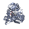
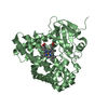
| Title | Crystal structure of Q4D6Q6, a conserved kinetoplastid-specific protein from Trypanosoma cruzi | ||||||
 Components Components | Uncharacterized protein | ||||||
 Keywords Keywords | UNKNOWN FUNCTION / Q4D6Q5 / Trypanosoma / neglected diseases | ||||||
| Function / homology | metal ion binding / IODIDE ION / : / Paraflagellar rod component Function and homology information Function and homology information | ||||||
| Biological species |  | ||||||
| Method |  X-RAY DIFFRACTION / SYNCHROTRON / SAD / Resolution: 1.47 Å X-RAY DIFFRACTION / SYNCHROTRON / SAD / Resolution: 1.47 Å | ||||||
 Authors Authors | Roske, Y. / Heinemann, U. | ||||||
| Funding support |  Brazil, 1items Brazil, 1items
| ||||||
 Citation Citation | Journal: J.Struct.Biol. / Year: 2020 Title: Crystal structure of Q4D6Q6, a conserved kinetoplastid-specific protein from Trypanosoma cruzi. Authors: D'Andrea, E.D. / Roske, Y. / Oliveira, G.A.P. / Cremer, N. / Diehl, A. / Schmieder, P. / Heinemann, U. / Oschkinat, H. / Pires, J.R. | ||||||
| History |
|
- Structure visualization
| Structure viewer | Molecule: MolmilJmol/JSmol |
|---|
- Downloads & links
-Download
| PDBx/mmCIF format | 6xyb.cif.gz | 195.4 KB | Display | PDBx/mmCIF format |
|---|---|---|---|---|
| PDB format | pdb6xyb.ent.gz | 154 KB | Display | PDB format |
| PDBx/mmJSON format | 6xyb.json.gz | Tree view | PDBx/mmJSON format | |
| Others |  Other downloads Other downloads |
-Validation report
| Summary document | 6xyb_validation.pdf.gz | 467.6 KB | Display | wwPDB validaton report |
|---|---|---|---|---|
| Full document | 6xyb_full_validation.pdf.gz | 472.4 KB | Display | |
| Data in XML | 6xyb_validation.xml.gz | 23.7 KB | Display | |
| Data in CIF | 6xyb_validation.cif.gz | 31.5 KB | Display | |
| Arichive directory | https://data.pdbj.org/pub/pdb/validation_reports/xy/6xybftp://data.pdbj.org/pub/pdb/validation_reports/xy/6xyb | HTTPS FTP |
-Related structure data


-Links
 PDBj
PDBj- Assembly
| Deposited unit | 
| |||||||||||||||||||||||||||||||||||||||||||||||||||||||||||||||||||||||||||||||||||||||||||||||||||||||||||||||||
|---|---|---|---|---|---|---|---|---|---|---|---|---|---|---|---|---|---|---|---|---|---|---|---|---|---|---|---|---|---|---|---|---|---|---|---|---|---|---|---|---|---|---|---|---|---|---|---|---|---|---|---|---|---|---|---|---|---|---|---|---|---|---|---|---|---|---|---|---|---|---|---|---|---|---|---|---|---|---|---|---|---|---|---|---|---|---|---|---|---|---|---|---|---|---|---|---|---|---|---|---|---|---|---|---|---|---|---|---|---|---|---|---|---|---|
| 1 |
| |||||||||||||||||||||||||||||||||||||||||||||||||||||||||||||||||||||||||||||||||||||||||||||||||||||||||||||||||
| Unit cell |
| |||||||||||||||||||||||||||||||||||||||||||||||||||||||||||||||||||||||||||||||||||||||||||||||||||||||||||||||||
| Noncrystallographic symmetry (NCS) | NCS domain:
NCS domain segments:
NCS oper:
|
-Components
-Protein , 1 types, 4 molecules ABCD
| #1: Protein | Mass: 13370.087 Da / Num. of mol.: 4 Source method: isolated from a genetically manipulated source Source: (gene. exp.) Gene: Tc00.1047053511733.90 / Plasmid: pET28a / Production host:  |
|---|
-Non-polymers , 5 types, 229 molecules 


| #2: Chemical |  Mass: 126.904 Da / Num. of mol.: 2 / Source method: obtained synthetically / Formula: I Mass: 126.904 Da / Num. of mol.: 2 / Source method: obtained synthetically / Formula: I#3: Chemical | ChemComp-K / |  Mass: 39.098 Da / Num. of mol.: 1 / Source method: obtained synthetically / Formula: K Mass: 39.098 Da / Num. of mol.: 1 / Source method: obtained synthetically / Formula: K#4: Chemical |  Mass: 24.305 Da / Num. of mol.: 2 / Source method: obtained synthetically / Formula: Mg Mass: 24.305 Da / Num. of mol.: 2 / Source method: obtained synthetically / Formula: Mg#5: Chemical | ChemComp-CL / |  Mass: 35.453 Da / Num. of mol.: 1 / Source method: obtained synthetically / Formula: Cl Mass: 35.453 Da / Num. of mol.: 1 / Source method: obtained synthetically / Formula: Cl#6: Water | ChemComp-HOH / | Mass: 18.015 Da / Num. of mol.: 223 / Source method: isolated from a natural source / Formula: H2O |
|---|
-Details
| Has ligand of interest | Y |
|---|---|
| Has protein modification | Y |
-Experimental details
-Experiment
| Experiment | Method: X-RAY DIFFRACTION / Number of used crystals: 1 |
|---|
- Sample preparation
| Crystal | Density Matthews: 1.91 Å3/Da / Density % sol: 35.5 % |
|---|---|
| Crystal grow | Temperature: 293 K / Method: vapor diffusion, sitting drop / Details: 20% PEG 5000 MME, 0.2 M potassium iodide |
-Data collection
| Diffraction | Mean temperature: 100 K / Serial crystal experiment: N |
|---|---|
| Diffraction source | Source: SYNCHROTRON / Site: BESSY  / Beamline: 14.1 / Wavelength: 0.91841 Å / Beamline: 14.1 / Wavelength: 0.91841 Å |
| Detector | Type: DECTRIS PILATUS3 6M / Detector: PIXEL / Date: Aug 20, 2014 |
| Radiation | Protocol: SINGLE WAVELENGTH / Monochromatic (M) / Laue (L): M / Scattering type: x-ray |
| Radiation wavelength | Wavelength: 0.91841 Å / Relative weight: 1 |
| Reflection | Resolution: 1.46→34.71 Å / Num. obs: 70953 / % possible obs: 99.4 % / Redundancy: 3.7 % / CC1/2: 0.998 / Rrim(I) all: 0.067 / Net I/σ(I): 10.8 |
| Reflection shell | Resolution: 1.46→1.55 Å / Redundancy: 3.5 % / Num. unique obs: 11262 / CC1/2: 0.642 / Rrim(I) all: 0.824 / % possible all: 98.2 |
- Processing
| Software |
| |||||||||||||||||||||||||||||||||||||||||||||||||||||||||||||||||||||||||||||||||||||||||||||||||||||||||||||||||||||||||||||
|---|---|---|---|---|---|---|---|---|---|---|---|---|---|---|---|---|---|---|---|---|---|---|---|---|---|---|---|---|---|---|---|---|---|---|---|---|---|---|---|---|---|---|---|---|---|---|---|---|---|---|---|---|---|---|---|---|---|---|---|---|---|---|---|---|---|---|---|---|---|---|---|---|---|---|---|---|---|---|---|---|---|---|---|---|---|---|---|---|---|---|---|---|---|---|---|---|---|---|---|---|---|---|---|---|---|---|---|---|---|---|---|---|---|---|---|---|---|---|---|---|---|---|---|---|---|---|
| Refinement | Method to determine structure: SAD / Resolution: 1.47→34.71 Å / Cor.coef. Fo:Fc: 0.976 / Cor.coef. Fo:Fc free: 0.965 / SU B: 1.374 / SU ML: 0.025 / Cross valid method: THROUGHOUT / σ(F): 0 / ESU R: 0.021 / ESU R Free: 0.017 / Stereochemistry target values: MAXIMUM LIKELIHOOD Details: HYDROGENS HAVE BEEN ADDED IN THE RIDING POSITIONS U VALUES : WITH TLS ADDED
| |||||||||||||||||||||||||||||||||||||||||||||||||||||||||||||||||||||||||||||||||||||||||||||||||||||||||||||||||||||||||||||
| Solvent computation | Ion probe radii: 0.8 Å / Shrinkage radii: 0.8 Å / VDW probe radii: 1.2 Å / Solvent model: MASK | |||||||||||||||||||||||||||||||||||||||||||||||||||||||||||||||||||||||||||||||||||||||||||||||||||||||||||||||||||||||||||||
| Displacement parameters | Biso max: 149.39 Å2 / Biso mean: 27.293 Å2 / Biso min: 7.86 Å2
| |||||||||||||||||||||||||||||||||||||||||||||||||||||||||||||||||||||||||||||||||||||||||||||||||||||||||||||||||||||||||||||
| Refinement step | Cycle: final / Resolution: 1.47→34.71 Å
| |||||||||||||||||||||||||||||||||||||||||||||||||||||||||||||||||||||||||||||||||||||||||||||||||||||||||||||||||||||||||||||
| Refine LS restraints |
| |||||||||||||||||||||||||||||||||||||||||||||||||||||||||||||||||||||||||||||||||||||||||||||||||||||||||||||||||||||||||||||
| Refine LS restraints NCS | Ens-ID: 1 / Number: 1265 / Refine-ID: X-RAY DIFFRACTION
| |||||||||||||||||||||||||||||||||||||||||||||||||||||||||||||||||||||||||||||||||||||||||||||||||||||||||||||||||||||||||||||
| LS refinement shell | Resolution: 1.47→1.506 Å / Rfactor Rfree error: 0
| |||||||||||||||||||||||||||||||||||||||||||||||||||||||||||||||||||||||||||||||||||||||||||||||||||||||||||||||||||||||||||||
| Refinement TLS params. | Method: refined / Refine-ID: X-RAY DIFFRACTION
| |||||||||||||||||||||||||||||||||||||||||||||||||||||||||||||||||||||||||||||||||||||||||||||||||||||||||||||||||||||||||||||
| Refinement TLS group |
|
