 Movie
Movie Controller

[English] 日本語
 Yorodumi
- PDB-6xo2: Structural Characterization of Beta Cyanoalanine Synthase from Te... -
+ Open data

- Basic information
| Entry | Database: PDB / ID: 6xo2 | ||||||
|---|---|---|---|---|---|---|---|
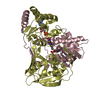
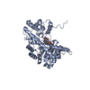
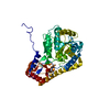
| Title | Structural Characterization of Beta Cyanoalanine Synthase from Tetranychus Urticae (two-spotted spider mite) | ||||||
 Components Components | Beta-cyanoalanine synthase | ||||||
 Keywords Keywords | BIOSYNTHETIC PROTEIN / Beta Cyanoalanine Synthase / PLP. two-spotted spider mite | ||||||
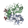
| Function / homology |  Function and homology information Function and homology information | ||||||
| Biological species |  Tetranychus urticae (two-spotted spider mite) Tetranychus urticae (two-spotted spider mite) | ||||||
| Method |  X-RAY DIFFRACTION / SYNCHROTRON / MOLECULAR REPLACEMENT / Resolution: 1.6 Å X-RAY DIFFRACTION / SYNCHROTRON / MOLECULAR REPLACEMENT / Resolution: 1.6 Å | ||||||
 Authors Authors | Daneshian, L. / Schlachter, C. / Dermauw, W. / Wybouw, N. / Van Leeuwen, T. / Chruszcz, M. | ||||||
| Funding support |  United States, 1items United States, 1items
| ||||||
 Citation Citation | Journal: Insect Biochem.Mol.Biol. / Year: 2022 Title: Structural and functional characterization of beta-cyanoalanine synthase from Tetranychus urticae. Authors: Daneshian, L. / Renggli, I. / Hanaway, R. / Offermann, L.R. / Schlachter, C.R. / Hernandez Arriaza, R. / Henry, S. / Prakash, R. / Wybouw, N. / Dermauw, W. / Shimizu, L.S. / Van Leeuwen, T. ...Authors: Daneshian, L. / Renggli, I. / Hanaway, R. / Offermann, L.R. / Schlachter, C.R. / Hernandez Arriaza, R. / Henry, S. / Prakash, R. / Wybouw, N. / Dermauw, W. / Shimizu, L.S. / Van Leeuwen, T. / Makris, T.M. / Grbic, V. / Grbic, M. / Chruszcz, M. | ||||||
| History |
|
- Structure visualization
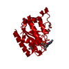
| Structure viewer | Molecule: MolmilJmol/JSmol |
|---|
- Downloads & links
-Download
| PDBx/mmCIF format | 6xo2.cif.gz | 130.6 KB | Display | PDBx/mmCIF format |
|---|---|---|---|---|
| PDB format | pdb6xo2.ent.gz | 99.2 KB | Display | PDB format |
| PDBx/mmJSON format | 6xo2.json.gz | Tree view | PDBx/mmJSON format | |
| Others |  Other downloads Other downloads |
-Validation report
| Arichive directory | https://data.pdbj.org/pub/pdb/validation_reports/xo/6xo2ftp://data.pdbj.org/pub/pdb/validation_reports/xo/6xo2 | HTTPS FTP |
|---|
-Related structure data
| Related structure data | 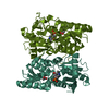 7mfjC 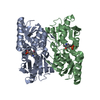 6pmuS S: Starting model for refinement C: citing same article ( |
|---|---|
| Similar structure data |

-Links
 PDBj
PDBj
- Assembly
| Deposited unit | 
| ||||||||||||
|---|---|---|---|---|---|---|---|---|---|---|---|---|---|
| 1 | 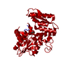
| ||||||||||||
| Unit cell |
| ||||||||||||
| Components on special symmetry positions |
|
-Components
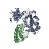
| #1: Protein | Mass: 34517.297 Da / Num. of mol.: 1 Source method: isolated from a genetically manipulated source Source: (gene. exp.) Tetranychus urticae (two-spotted spider mite)Gene: 107363798 / Plasmid: pMCSG35 / Production host:  |
|---|---|
| #2: Chemical | ChemComp-PLP /   Mass: 247.142 Da / Num. of mol.: 1 / Source method: obtained synthetically / Formula: C8H10NO6P / Feature type: SUBJECT OF INVESTIGATION Mass: 247.142 Da / Num. of mol.: 1 / Source method: obtained synthetically / Formula: C8H10NO6P / Feature type: SUBJECT OF INVESTIGATION |
| #3: Chemical | ChemComp-ACT /   Mass: 59.044 Da / Num. of mol.: 1 / Source method: obtained synthetically / Formula: C2H3O2 Mass: 59.044 Da / Num. of mol.: 1 / Source method: obtained synthetically / Formula: C2H3O2 |
| #4: Water | ChemComp-HOH /  Mass: 18.015 Da / Num. of mol.: 290 / Source method: isolated from a natural source / Formula: H2O Mass: 18.015 Da / Num. of mol.: 290 / Source method: isolated from a natural source / Formula: H2O |
| Has ligand of interest | Y |
-Experimental details
-Experiment
| Experiment | Method: X-RAY DIFFRACTION / Number of used crystals: 1 |
|---|
- Sample preparation
| Crystal | Density Matthews: 2.15 Å3/Da / Density % sol: 42.89 % |
|---|---|
| Crystal grow | Temperature: 277 K / Method: vapor diffusion, sitting drop / pH: 7 / Details: 0.2 M Ammonium Chloride, 12% PEG 3350, pH 7.0. |
-Data collection
| Diffraction | Mean temperature: 100 K / Serial crystal experiment: N |
|---|---|
| Diffraction source | Source: SYNCHROTRON / Site: APS / Beamline: 22-ID / Wavelength: 1 Å |
| Detector | Type: DECTRIS EIGER X 16M / Detector: PIXEL / Date: Jun 10, 2018 |
| Radiation | Protocol: SINGLE WAVELENGTH / Monochromatic (M) / Laue (L): M / Scattering type: x-ray |
| Radiation wavelength | Wavelength: 1 Å / Relative weight: 1 |
| Reflection | Resolution: 1.6→40 Å / Num. obs: 40447 / % possible obs: 99.4 % / Redundancy: 6.3 % / Rmerge(I) obs: 0.079 / Rpim(I) all: 0.046 / Rrim(I) all: 0.087 / Rsym value: 0.079 / Net I/av σ(I): 33.5 / Net I/σ(I): 41.05 |
| Reflection shell | Resolution: 1.6→1.63 Å / Redundancy: 4.8 % / Rmerge(I) obs: 0.681 / Mean I/σ(I) obs: 2.1 / Num. unique obs: 1971 / CC1/2: 0.757 / CC star: 0.928 / Rpim(I) all: 0.335 / Rrim(I) all: 0.763 / Rsym value: 0.681 / % possible all: 99.4 |
- Processing
| Software |
| ||||||||||||||||||||||||||||||||||||||||||||||||||||||||||||||||||||||||||||||||||||||||||||||||||||
|---|---|---|---|---|---|---|---|---|---|---|---|---|---|---|---|---|---|---|---|---|---|---|---|---|---|---|---|---|---|---|---|---|---|---|---|---|---|---|---|---|---|---|---|---|---|---|---|---|---|---|---|---|---|---|---|---|---|---|---|---|---|---|---|---|---|---|---|---|---|---|---|---|---|---|---|---|---|---|---|---|---|---|---|---|---|---|---|---|---|---|---|---|---|---|---|---|---|---|---|---|---|
| Refinement | Method to determine structure: MOLECULAR REPLACEMENT Starting model: 6pmu Resolution: 1.6→38.47 Å / Cor.coef. Fo:Fc: 0.971 / Cor.coef. Fo:Fc free: 0.964 / SU B: 3.522 / SU ML: 0.058 / Cross valid method: THROUGHOUT / σ(F): 0 / ESU R: 0.08 / ESU R Free: 0.079 / Stereochemistry target values: MAXIMUM LIKELIHOOD Details: HYDROGENS HAVE BEEN ADDED IN THE RIDING POSITIONS U VALUES : WITH TLS ADDED
| ||||||||||||||||||||||||||||||||||||||||||||||||||||||||||||||||||||||||||||||||||||||||||||||||||||
| Solvent computation | Ion probe radii: 0.8 Å / Shrinkage radii: 0.8 Å / VDW probe radii: 1.2 Å / Solvent model: BABINET MODEL WITH MASK | ||||||||||||||||||||||||||||||||||||||||||||||||||||||||||||||||||||||||||||||||||||||||||||||||||||
| Displacement parameters | Biso max: 103.15 Å2 / Biso mean: 29.786 Å2 / Biso min: 18.69 Å2
| ||||||||||||||||||||||||||||||||||||||||||||||||||||||||||||||||||||||||||||||||||||||||||||||||||||
| Refinement step | Cycle: final / Resolution: 1.6→38.47 Å
| ||||||||||||||||||||||||||||||||||||||||||||||||||||||||||||||||||||||||||||||||||||||||||||||||||||
| Refine LS restraints |
| ||||||||||||||||||||||||||||||||||||||||||||||||||||||||||||||||||||||||||||||||||||||||||||||||||||
| LS refinement shell | Resolution: 1.601→1.642 Å / Rfactor Rfree error: 0 / Total num. of bins used: 20
| ||||||||||||||||||||||||||||||||||||||||||||||||||||||||||||||||||||||||||||||||||||||||||||||||||||
| Refinement TLS params. | Method: refined / Refine-ID: X-RAY DIFFRACTION
| ||||||||||||||||||||||||||||||||||||||||||||||||||||||||||||||||||||||||||||||||||||||||||||||||||||
| Refinement TLS group |
|
