 Movie
Movie Controller

[English] 日本語
 Yorodumi
- PDB-3cls: Crystal structure of the R236C mutant of ETF from Methylophilus m... -
+ Open data

- Basic information
| Entry | Database: PDB / ID: 3cls | ||||||
|---|---|---|---|---|---|---|---|
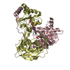
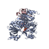
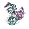
| Title | Crystal structure of the R236C mutant of ETF from Methylophilus methylotrophus | ||||||
 Components Components |
| ||||||
 Keywords Keywords | ELECTRON TRANSPORT / ETF / TMADH / electron transfer / dynamic interface / FAD / Flavoprotein / Transport | ||||||
| Function / homology |  Function and homology information Function and homology informationfatty acid beta-oxidation using acyl-CoA dehydrogenase / flavin adenine dinucleotide binding / electron transfer activity / nucleotide binding Similarity search - Function | ||||||
| Biological species |  Methylophilus methylotrophus (bacteria) Methylophilus methylotrophus (bacteria) | ||||||
| Method |  X-RAY DIFFRACTION / SYNCHROTRON / FOURIER SYNTHESIS / Resolution: 1.65 Å X-RAY DIFFRACTION / SYNCHROTRON / FOURIER SYNTHESIS / Resolution: 1.65 Å | ||||||
 Authors Authors | Katona, G. / Leys, D. | ||||||
 Citation Citation | Journal: Biochemistry / Year: 2008 Title: Probing the dynamic interface between trimethylamine dehydrogenase (TMADH) and electron transferring flavoprotein (ETF) in the TMADH-2ETF complex: role of the Arg-alpha237 (ETF) and Tyr-442 (TMADH) residue pair. Authors: Burgess, S.G. / Messiha, H.L. / Katona, G. / Rigby, S.E. / Leys, D. / Scrutton, N.S. | ||||||
| History |
|
- Structure visualization
| Structure viewer | Molecule: MolmilJmol/JSmol |
|---|
- Downloads & links
-Download
| PDBx/mmCIF format | 3cls.cif.gz | 246.3 KB | Display | PDBx/mmCIF format |
|---|---|---|---|---|
| PDB format | pdb3cls.ent.gz | 193.5 KB | Display | PDB format |
| PDBx/mmJSON format | 3cls.json.gz | Tree view | PDBx/mmJSON format | |
| Others |  Other downloads Other downloads |
-Validation report
| Arichive directory | https://data.pdbj.org/pub/pdb/validation_reports/cl/3clsftp://data.pdbj.org/pub/pdb/validation_reports/cl/3cls | HTTPS FTP |
|---|
-Related structure data
| Related structure data |  3clrC  3cltC  3cluC  1o97S S: Starting model for refinement C: citing same article ( |
|---|---|
| Similar structure data |


-Links
 PDBj
PDBj


- Assembly
| Deposited unit | 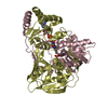
| ||||||||
|---|---|---|---|---|---|---|---|---|---|
| 1 |
| ||||||||
| Unit cell |
|
-Components
| #1: Protein | Mass: 28929.850 Da / Num. of mol.: 1 / Mutation: R237C Source method: isolated from a genetically manipulated source Source: (gene. exp.) Methylophilus methylotrophus (bacteria)Gene: etfB / Production host: |
|---|---|
| #2: Protein | Mass: 33700.090 Da / Num. of mol.: 1 Source method: isolated from a genetically manipulated source Source: (gene. exp.) Methylophilus methylotrophus (bacteria)Gene: etfA / Production host: |
| #3: Chemical | ChemComp-AMP /   Mass: 347.221 Da / Num. of mol.: 1 / Source method: obtained synthetically / Formula: C10H14N5O7P / Comment: AMP*YM Mass: 347.221 Da / Num. of mol.: 1 / Source method: obtained synthetically / Formula: C10H14N5O7P / Comment: AMP*YM |
| #4: Chemical | ChemComp-FAD /   Mass: 785.550 Da / Num. of mol.: 1 / Source method: obtained synthetically / Formula: C27H33N9O15P2 / Comment: FAD*YM Mass: 785.550 Da / Num. of mol.: 1 / Source method: obtained synthetically / Formula: C27H33N9O15P2 / Comment: FAD*YM |
| #5: Water | ChemComp-HOH /  Mass: 18.015 Da / Num. of mol.: 693 / Source method: isolated from a natural source / Formula: H2O Mass: 18.015 Da / Num. of mol.: 693 / Source method: isolated from a natural source / Formula: H2O |
-Experimental details
-Experiment
| Experiment | Method: X-RAY DIFFRACTION / Number of used crystals: 1 |
|---|
- Sample preparation
| Crystal | Density Matthews: 2.7 Å3/Da / Density % sol: 54.46 % |
|---|---|
| Crystal grow | Method: vapor diffusion, hanging drop / pH: 6.2 Details: limited digest with trypsin, 2.0 M sodium phosphate, pH 6.2, VAPOR DIFFUSION, HANGING DROP |
-Data collection
| Diffraction | Mean temperature: 100 K |
|---|---|
| Diffraction source | Source: SYNCHROTRON / Site: ESRF  / Beamline: ID14-1 / Wavelength: 0.933 / Beamline: ID14-1 / Wavelength: 0.933 |
| Detector | Type: ADSC QUANTUM 210 / Detector: CCD / Date: Apr 1, 2005 |
| Radiation | Protocol: SINGLE WAVELENGTH / Monochromatic (M) / Laue (L): M / Scattering type: x-ray |
| Radiation wavelength | Wavelength: 0.933 Å / Relative weight: 1 |
| Reflection | Resolution: 1.65→20 Å / Num. all: 72327 / Num. obs: 72327 / % possible obs: 95.6 % / Observed criterion σ(F): 0 / Observed criterion σ(I): 0 / Redundancy: 3.8 % / Rsym value: 0.084 / Net I/σ(I): 7.3 |
- Processing
| Software |
| ||||||||||||||||||||||||||||||||||||||||||||||||||||||||||||||||||||||||||||||||||||||||||||||||||||||||||||||||||||||||||||||||||||||||||||||||||||||||||||||||||||||||||
|---|---|---|---|---|---|---|---|---|---|---|---|---|---|---|---|---|---|---|---|---|---|---|---|---|---|---|---|---|---|---|---|---|---|---|---|---|---|---|---|---|---|---|---|---|---|---|---|---|---|---|---|---|---|---|---|---|---|---|---|---|---|---|---|---|---|---|---|---|---|---|---|---|---|---|---|---|---|---|---|---|---|---|---|---|---|---|---|---|---|---|---|---|---|---|---|---|---|---|---|---|---|---|---|---|---|---|---|---|---|---|---|---|---|---|---|---|---|---|---|---|---|---|---|---|---|---|---|---|---|---|---|---|---|---|---|---|---|---|---|---|---|---|---|---|---|---|---|---|---|---|---|---|---|---|---|---|---|---|---|---|---|---|---|---|---|---|---|---|---|---|---|
| Refinement | Method to determine structure: FOURIER SYNTHESIS Starting model: 1O97 Resolution: 1.65→20 Å / Cor.coef. Fo:Fc: 0.973 / Cor.coef. Fo:Fc free: 0.963 / SU B: 3.461 / SU ML: 0.065 / Cross valid method: THROUGHOUT / ESU R: 0.087 / ESU R Free: 0.086 / Stereochemistry target values: MAXIMUM LIKELIHOOD / Details: HYDROGENS HAVE BEEN ADDED IN THE RIDING POSITIONS
| ||||||||||||||||||||||||||||||||||||||||||||||||||||||||||||||||||||||||||||||||||||||||||||||||||||||||||||||||||||||||||||||||||||||||||||||||||||||||||||||||||||||||||
| Solvent computation | Ion probe radii: 0.8 Å / Shrinkage radii: 0.8 Å / VDW probe radii: 1.2 Å / Solvent model: MASK | ||||||||||||||||||||||||||||||||||||||||||||||||||||||||||||||||||||||||||||||||||||||||||||||||||||||||||||||||||||||||||||||||||||||||||||||||||||||||||||||||||||||||||
| Displacement parameters | Biso mean: 14.334 Å2
| ||||||||||||||||||||||||||||||||||||||||||||||||||||||||||||||||||||||||||||||||||||||||||||||||||||||||||||||||||||||||||||||||||||||||||||||||||||||||||||||||||||||||||
| Refinement step | Cycle: LAST / Resolution: 1.65→20 Å
| ||||||||||||||||||||||||||||||||||||||||||||||||||||||||||||||||||||||||||||||||||||||||||||||||||||||||||||||||||||||||||||||||||||||||||||||||||||||||||||||||||||||||||
| Refine LS restraints |
| ||||||||||||||||||||||||||||||||||||||||||||||||||||||||||||||||||||||||||||||||||||||||||||||||||||||||||||||||||||||||||||||||||||||||||||||||||||||||||||||||||||||||||
| LS refinement shell | Resolution: 1.652→1.695 Å / Total num. of bins used: 20
| ||||||||||||||||||||||||||||||||||||||||||||||||||||||||||||||||||||||||||||||||||||||||||||||||||||||||||||||||||||||||||||||||||||||||||||||||||||||||||||||||||||||||||
| Refinement TLS params. | Method: refined / Refine-ID: X-RAY DIFFRACTION
| ||||||||||||||||||||||||||||||||||||||||||||||||||||||||||||||||||||||||||||||||||||||||||||||||||||||||||||||||||||||||||||||||||||||||||||||||||||||||||||||||||||||||||
| Refinement TLS group |
|
