| Entry | Database: PDB / ID: 6xn8
|
|---|
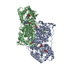
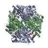
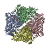
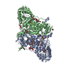
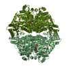
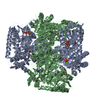
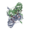
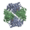
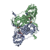
| Title | Crystal Structure of 2-hydroxyacyl CoA lyase (HACL) from Rhodospirillales bacterium URHD0017 |
|---|
 Components Components | 2-hydroxyacyl-CoA lyase 1 |
|---|
 Keywords Keywords | LYASE / 2-hydroxyacyl CoA lyase / HACL / acyloin condensation / thiamine pyrophosphate |
|---|
| Function / homology |  Function and homology information Function and homology information
fatty acid alpha-oxidation / thiamine pyrophosphate binding / peroxisome / lyase activity / nucleotide binding / magnesium ion bindingSimilarity search - Function TPP-binding domain containing protein HACL1-like / TPP-binding enzyme, conserved site / Thiamine pyrophosphate enzymes signature. / Thiamine pyrophosphate enzyme, central domain / Thiamine pyrophosphate enzyme, central domain / Thiamine pyrophosphate enzyme, N-terminal TPP-binding domain / Thiamine pyrophosphate enzyme, N-terminal TPP binding domain / Thiamine pyrophosphate enzyme, C-terminal TPP-binding / Thiamine pyrophosphate enzyme, C-terminal TPP binding domain / Thiamin diphosphate-binding fold / DHS-like NAD/FAD-binding domain superfamilySimilarity search - Domain/homology |
|---|
| Biological species |  Rhodospirillales bacterium URHD0017 (bacteria) Rhodospirillales bacterium URHD0017 (bacteria) |
|---|
| Method |  X-RAY DIFFRACTION / SYNCHROTRON / MOLECULAR REPLACEMENT / molecular replacement / Resolution: 1.95 Å X-RAY DIFFRACTION / SYNCHROTRON / MOLECULAR REPLACEMENT / molecular replacement / Resolution: 1.95 Å |
|---|
 Authors Authors | Miller, M.D. / Xu, W. / Olmos Jr., J.L. / Chou, A. / Clomburg, J.M. / Gonzalez, R. / Philips Jr., G.N. |
|---|
| Funding support |  United States, 5items United States, 5items | Organization | Grant number | Country |
|---|
| National Science Foundation (NSF, United States) | STC 1231306 | United States | | National Science Foundation (NSF, United States) | CBET-1605999 | United States | | National Institutes of Health/National Institute of General Medical Sciences (NIH/NIGMS) | R01 GM115261 | United States | | National Institutes of Health/National Cancer Institute (NIH/NCI) | R01 CA217255 | United States | | National Science Foundation (NSF, United States) | GRFP 1450681 | United States |
|
|---|
 Citation Citation | Journal: To Be Published
Title: Crystal Structure of 2-hydroxyacyl CoA lyase (HACL) from Rhodospirillales bacterium URHD0017
Authors: Chou, A. / Miller, M.D. / Olmos Jr., J.L. / Xu, W. / Clomburg, J.M. / Gonzalez, R. / Philips Jr., G.N. |
|---|
| History | | Deposition | Jul 2, 2020 | Deposition site: RCSB / Processing site: RCSB |
|---|
| Revision 1.0 | Jul 7, 2021 | Provider: repository / Type: Initial release |
|---|
| Revision 1.1 | Oct 18, 2023 | Group: Data collection / Database references / Refinement description
Category: chem_comp_atom / chem_comp_bond ...chem_comp_atom / chem_comp_bond / database_2 / pdbx_initial_refinement_model / struct_ncs_dom_lim
Item: _database_2.pdbx_DOI / _database_2.pdbx_database_accession ..._database_2.pdbx_DOI / _database_2.pdbx_database_accession / _struct_ncs_dom_lim.beg_auth_comp_id / _struct_ncs_dom_lim.beg_label_asym_id / _struct_ncs_dom_lim.beg_label_comp_id / _struct_ncs_dom_lim.beg_label_seq_id / _struct_ncs_dom_lim.end_auth_comp_id / _struct_ncs_dom_lim.end_label_asym_id / _struct_ncs_dom_lim.end_label_comp_id / _struct_ncs_dom_lim.end_label_seq_id |
|---|
|
|---|
 Movie
Movie Controller

 Yorodumi
 Open data
 Basic information
 Structure visualization
 Downloads & links
 Other downloads
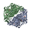


 PDBj
PDBj


 Assembly