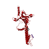
 ムービー
ムービー コントローラー


+ データを開く

- 基本情報
基本情報
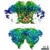
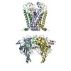
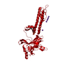
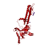
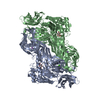
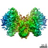
| 登録情報 | データベース: PDB / ID: 6xit | ||||||
|---|---|---|---|---|---|---|---|
| タイトル | Cryo-EM structure of the G protein-gated inward rectifier K+ channel GIRK2 (Kir3.2) in complex with PIP2 | ||||||
 要素 要素 | G protein-activated inward rectifier potassium channel 2 | ||||||
 キーワード キーワード | MEMBRANE PROTEIN / G protein-coupled inwardly rectifying potassium channels / PIP2 | ||||||
| 機能・相同性 |  機能・相同性情報 機能・相同性情報G-protein activated inward rectifier potassium channel activity / Activation of G protein gated Potassium channels / Inhibition of voltage gated Ca2+ channels via Gbeta/gamma subunits / inward rectifier potassium channel complex / voltage-gated monoatomic ion channel activity involved in regulation of presynaptic membrane potential / inward rectifier potassium channel activity / regulation of monoatomic ion transmembrane transport / neuronal cell body membrane / potassium ion import across plasma membrane / parallel fiber to Purkinje cell synapse ...G-protein activated inward rectifier potassium channel activity / Activation of G protein gated Potassium channels / Inhibition of voltage gated Ca2+ channels via Gbeta/gamma subunits / inward rectifier potassium channel complex / voltage-gated monoatomic ion channel activity involved in regulation of presynaptic membrane potential / inward rectifier potassium channel activity / regulation of monoatomic ion transmembrane transport / neuronal cell body membrane / potassium ion import across plasma membrane / parallel fiber to Purkinje cell synapse / G-protein alpha-subunit binding / potassium channel activity / negative regulation of insulin secretion / presynapse / presynaptic membrane / postsynapse / axon / dendrite / cell surface / membrane / plasma membrane 類似検索 - 分子機能 | ||||||
| 生物種 |  | ||||||
| 手法 | 電子顕微鏡法 / 単粒子再構成法 / クライオ電子顕微鏡法 / 解像度: 3.3 Å | ||||||
 データ登録者 データ登録者 | Niu, Y. / Tao, X. / MacKinnon, R. | ||||||
| 資金援助 |  米国, 1件 米国, 1件
| ||||||
 引用 引用 | ジャーナル: Elife / 年: 2020 タイトル: Cryo-EM analysis of PIP regulation in mammalian GIRK channels. 著者: Yiming Niu / Xiao Tao / Kouki K Touhara / Roderick MacKinnon / 要旨: G-protein-gated inward rectifier potassium (GIRK) channels are regulated by G proteins and PIP. Here, using cryo-EM single particle analysis we describe the equilibrium ensemble of structures of ...G-protein-gated inward rectifier potassium (GIRK) channels are regulated by G proteins and PIP. Here, using cryo-EM single particle analysis we describe the equilibrium ensemble of structures of neuronal GIRK2 as a function of the C8-PIP concentration. We find that PIP shifts the equilibrium between two distinguishable structures of neuronal GIRK (GIRK2), extended and docked, towards the docked form. In the docked form the cytoplasmic domain, to which G binds, becomes accessible to the cytoplasmic membrane surface where G resides. Furthermore, PIP binding reshapes the G binding surface on the cytoplasmic domain, preparing it to receive G. We find that cardiac GIRK (GIRK1/4) can also exist in both extended and docked conformations. These findings lead us to conclude that PIP influences GIRK channels in a structurally similar manner to Kir2.2 channels. In Kir2.2 channels, the PIP-induced conformational changes open the pore. In GIRK channels, they prepare the channel for activation by G. | ||||||
| 履歴 |
|
- 構造の表示
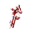
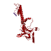
構造の表示
| ムービー |
ムービービューア |
|---|---|
| 構造ビューア | 分子: MolmilJmol/JSmol |
- ダウンロードとリンク
-ダウンロード
| PDBx/mmCIF形式 | 6xit.cif.gz | 312.2 KB | 表示 | PDBx/mmCIF形式 |
|---|---|---|---|---|
| PDB形式 | pdb6xit.ent.gz | 237 KB | 表示 | PDB形式 |
| PDBx/mmJSON形式 | 6xit.json.gz | ツリー表示 | PDBx/mmJSON形式 | |
| その他 |  その他のダウンロード その他のダウンロード |
-検証レポート
| アーカイブディレクトリ | https://data.pdbj.org/pub/pdb/validation_reports/xi/6xitftp://data.pdbj.org/pub/pdb/validation_reports/xi/6xit | HTTPS FTP |
|---|
-関連構造データ


-リンク
 PDBj
PDBj
- 集合体
集合体
| 登録構造単位 | 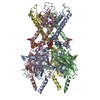
|
|---|---|
| 1 |
|
-要素
| #1: タンパク質 | 分子量: 39061.957 Da / 分子数: 4 / 由来タイプ: 組換発現 / 由来: (組換発現)  Komagataella pastoris (菌類) / 参照: UniProt: A0A338P6L0, UniProt: P48542*PLUS Komagataella pastoris (菌類) / 参照: UniProt: A0A338P6L0, UniProt: P48542*PLUS#2: 化合物 | ChemComp-PIO / [(   分子量: 746.566 Da / 分子数: 4 / 由来タイプ: 合成 / 式: C25H49O19P3 / タイプ: SUBJECT OF INVESTIGATION 分子量: 746.566 Da / 分子数: 4 / 由来タイプ: 合成 / 式: C25H49O19P3 / タイプ: SUBJECT OF INVESTIGATION#3: 化合物 |   分子量: 39.098 Da / 分子数: 3 / 由来タイプ: 合成 / 式: K / タイプ: SUBJECT OF INVESTIGATION 分子量: 39.098 Da / 分子数: 3 / 由来タイプ: 合成 / 式: K / タイプ: SUBJECT OF INVESTIGATION研究の焦点であるリガンドがあるか | Y | Has protein modification | Y | |
|---|
-実験情報
-実験
| 実験 | 手法: 電子顕微鏡法 |
|---|---|
| EM実験 | 試料の集合状態: PARTICLE / 3次元再構成法: 単粒子再構成法 |
- 試料調製
試料調製
| 構成要素 | 名称: Homo-tetrameric assembly of G protein-activated inward rectifier potassium channel 2 タイプ: COMPLEX / Entity ID: #1 / 由来: RECOMBINANT |
|---|---|
| 分子量 | 実験値: NO |
| 由来(天然) | 生物種: |
| 由来(組換発現) | 生物種: Komagataella pastoris (菌類) |
| 緩衝液 | pH: 7.5 詳細: 20 mM Tris-HCl pH 7.5, 150 mM KCl, 10 mM DTT, 1 mM DETA, 0.2% DM and 0.01% CHS |
| 試料 | 濃度: 6 mg/ml / 包埋: NO / シャドウイング: NO / 染色: NO / 凍結: YES |
| 試料支持 | グリッドの材料: GOLD / グリッドのサイズ: 400 divisions/in. / グリッドのタイプ: Quantifoil R1.2/1.3 |
| 急速凍結 | 装置: FEI VITROBOT MARK IV / 凍結剤: ETHANE / 湿度: 95 % / 凍結前の試料温度: 298 K |
- 電子顕微鏡撮影
| 実験機器 |  モデル: Titan Krios / 画像提供: FEI Company |
|---|---|
| 顕微鏡 | モデル: FEI TITAN KRIOS |
| 電子銃 | 電子線源:  FIELD EMISSION GUN / 加速電圧: 300 kV / 照射モード: FLOOD BEAM FIELD EMISSION GUN / 加速電圧: 300 kV / 照射モード: FLOOD BEAM |
| 電子レンズ | モード: BRIGHT FIELD / C2レンズ絞り径: 100 µm |
| 試料ホルダ | 凍結剤: NITROGEN 試料ホルダーモデル: FEI TITAN KRIOS AUTOGRID HOLDER |
| 撮影 | 平均露光時間: 10 sec. / 電子線照射量: 8 e/Å2 / 検出モード: SUPER-RESOLUTION フィルム・検出器のモデル: GATAN K2 SUMMIT (4k x 4k) 撮影したグリッド数: 1 / 実像数: 11349 |
| 画像スキャン | 動画フレーム数/画像: 50 / 利用したフレーム数/画像: 1-50 |
- 解析
解析
| EMソフトウェア |
| ||||||||||||||||||||||||||||||||||||||||||||||||
|---|---|---|---|---|---|---|---|---|---|---|---|---|---|---|---|---|---|---|---|---|---|---|---|---|---|---|---|---|---|---|---|---|---|---|---|---|---|---|---|---|---|---|---|---|---|---|---|---|---|
| CTF補正 | タイプ: NONE | ||||||||||||||||||||||||||||||||||||||||||||||||
| 対称性 | 点対称性: C4 (4回回転対称) | ||||||||||||||||||||||||||||||||||||||||||||||||
| 3次元再構成 | 解像度: 3.3 Å / 解像度の算出法: FSC 0.143 CUT-OFF / 粒子像の数: 155128 / 対称性のタイプ: POINT | ||||||||||||||||||||||||||||||||||||||||||||||||
| 原子モデル構築 | PDB-ID: 3SYA PDB chain-ID: A / Accession code: 3SYA / Pdb chain residue range: 53-380 / Source name: PDB / タイプ: experimental model |

