 Movie
Movie Controller

[English] 日本語
 Yorodumi
- EMDB-22201: Cryo-EM structure of the G protein-gated inward rectifier K+ chan... -
+ Open data

- Basic information
| Entry | Database: EMDB / ID: EMD-22201 | |||||||||
|---|---|---|---|---|---|---|---|---|---|---|
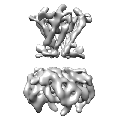
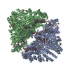
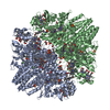
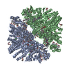
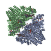
| Title | Cryo-EM structure of the G protein-gated inward rectifier K+ channel GIRK1/4 (Kir3.1/Kir3.4) in apo form | |||||||||
 Map data Map data | Cryo-EM structure of the G protein-gated inward rectifier K+ channel GIRK1/4 (Kir3.1/Kir3.4) in apo form | |||||||||
 Sample Sample |
| |||||||||
| Function / homology |  Function and homology information Function and homology informationventricular cardiac muscle cell membrane repolarization / G-protein activated inward rectifier potassium channel activity / voltage-gated potassium channel activity involved in atrial cardiac muscle cell action potential repolarization / inward rectifier potassium channel complex / membrane repolarization during atrial cardiac muscle cell action potential / voltage-gated potassium channel activity involved in ventricular cardiac muscle cell action potential repolarization / voltage-gated monoatomic ion channel activity involved in regulation of presynaptic membrane potential / inward rectifier potassium channel activity / potassium ion import across plasma membrane / parallel fiber to Purkinje cell synapse ...ventricular cardiac muscle cell membrane repolarization / G-protein activated inward rectifier potassium channel activity / voltage-gated potassium channel activity involved in atrial cardiac muscle cell action potential repolarization / inward rectifier potassium channel complex / membrane repolarization during atrial cardiac muscle cell action potential / voltage-gated potassium channel activity involved in ventricular cardiac muscle cell action potential repolarization / voltage-gated monoatomic ion channel activity involved in regulation of presynaptic membrane potential / inward rectifier potassium channel activity / potassium ion import across plasma membrane / parallel fiber to Purkinje cell synapse / regulation of heart rate by cardiac conduction / response to electrical stimulus / phosphatidylinositol-4,5-bisphosphate binding / voltage-gated potassium channel complex / T-tubule / potassium ion transmembrane transport / potassium ion transport / Activation of G protein gated Potassium channels / Inhibition of voltage gated Ca2+ channels via Gbeta/gamma subunits / presynaptic membrane / external side of plasma membrane / plasma membrane Similarity search - Function | |||||||||
| Biological species |  Homo sapiens (human) Homo sapiens (human) | |||||||||
| Method | single particle reconstruction / cryo EM / Resolution: 7.9 Å | |||||||||
 Authors Authors | Niu Y / Tao X / MacKinnon R | |||||||||
| Funding support |  United States, 1 items United States, 1 items
| |||||||||
 Citation Citation | Journal: Elife / Year: 2020 Title: Cryo-EM analysis of PIP regulation in mammalian GIRK channels. Authors: Yiming Niu / Xiao Tao / Kouki K Touhara / Roderick MacKinnon / Abstract: G-protein-gated inward rectifier potassium (GIRK) channels are regulated by G proteins and PIP. Here, using cryo-EM single particle analysis we describe the equilibrium ensemble of structures of ...G-protein-gated inward rectifier potassium (GIRK) channels are regulated by G proteins and PIP. Here, using cryo-EM single particle analysis we describe the equilibrium ensemble of structures of neuronal GIRK2 as a function of the C8-PIP concentration. We find that PIP shifts the equilibrium between two distinguishable structures of neuronal GIRK (GIRK2), extended and docked, towards the docked form. In the docked form the cytoplasmic domain, to which G binds, becomes accessible to the cytoplasmic membrane surface where G resides. Furthermore, PIP binding reshapes the G binding surface on the cytoplasmic domain, preparing it to receive G. We find that cardiac GIRK (GIRK1/4) can also exist in both extended and docked conformations. These findings lead us to conclude that PIP influences GIRK channels in a structurally similar manner to Kir2.2 channels. In Kir2.2 channels, the PIP-induced conformational changes open the pore. In GIRK channels, they prepare the channel for activation by G. | |||||||||
| History |
|
- Structure visualization
| Movie |
Movie viewer |
|---|---|
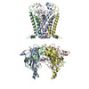
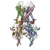
| Structure viewer | EM map: SurfViewMolmilJmol/JSmol |
| Supplemental images |
- Downloads & links
-EMDB archive
| Map data | emd_22201.map.gz | 58 MB | EMDB map data format | |
|---|---|---|---|---|
| Header (meta data) | emd-22201-v30.xmlemd-22201.xml | 13.4 KB 13.4 KB | Display Display | EMDB header |
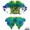
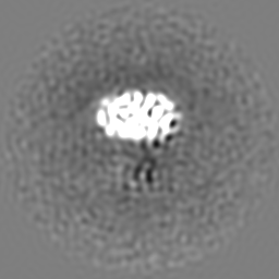
| Images |  emd_22201.png emd_22201.png | 51 KB | ||
| Archive directory |  http://ftp.pdbj.org/pub/emdb/structures/EMD-22201ftp://ftp.pdbj.org/pub/emdb/structures/EMD-22201 http://ftp.pdbj.org/pub/emdb/structures/EMD-22201ftp://ftp.pdbj.org/pub/emdb/structures/EMD-22201 | HTTPS FTP |
-Related structure data


-Links
| EMDB pages | EMDB (EBI/PDBe) / EMDataResource |
|---|
-Map
| File | Download / File: emd_22201.map.gz / Format: CCP4 / Size: 64 MB / Type: IMAGE STORED AS FLOATING POINT NUMBER (4 BYTES) | ||||||||||||||||||||||||||||||||||||||||||||||||||||||||||||||||||||
|---|---|---|---|---|---|---|---|---|---|---|---|---|---|---|---|---|---|---|---|---|---|---|---|---|---|---|---|---|---|---|---|---|---|---|---|---|---|---|---|---|---|---|---|---|---|---|---|---|---|---|---|---|---|---|---|---|---|---|---|---|---|---|---|---|---|---|---|---|---|
| Annotation | Cryo-EM structure of the G protein-gated inward rectifier K+ channel GIRK1/4 (Kir3.1/Kir3.4) in apo form | ||||||||||||||||||||||||||||||||||||||||||||||||||||||||||||||||||||
| Projections & slices | Image control
Images are generated by Spider. | ||||||||||||||||||||||||||||||||||||||||||||||||||||||||||||||||||||
| Voxel size | X=Y=Z: 1.03 Å | ||||||||||||||||||||||||||||||||||||||||||||||||||||||||||||||||||||
| Density |
| ||||||||||||||||||||||||||||||||||||||||||||||||||||||||||||||||||||
| Symmetry | Space group: 1 | ||||||||||||||||||||||||||||||||||||||||||||||||||||||||||||||||||||
| Details | EMDB XML:
CCP4 map header:
| ||||||||||||||||||||||||||||||||||||||||||||||||||||||||||||||||||||
 Z (Sec.)
 Y (Row.)
 X (Col.)


-Supplemental data
- Sample components
-Entire : Hetero-tetrameric assembly of G protein-activated inward rectifie...
| Entire | Name: Hetero-tetrameric assembly of G protein-activated inward rectifier potassium channel 1/4 |
|---|---|
| Components |
|
-Supramolecule #1: Hetero-tetrameric assembly of G protein-activated inward rectifie...
| Supramolecule | Name: Hetero-tetrameric assembly of G protein-activated inward rectifier potassium channel 1/4 type: complex / ID: 1 / Parent: 0 / Macromolecule list: all |
|---|---|
| Source (natural) | Organism: Homo sapiens (human) |
| Recombinant expression | Organism: Homo sapiens (human) / Recombinant strain: HEK293T |
-Macromolecule #1: G protein-activated inward rectifier potassium channel 1
| Macromolecule | Name: G protein-activated inward rectifier potassium channel 1 type: protein_or_peptide / ID: 1 / Enantiomer: LEVO |
|---|---|
| Source (natural) | Organism: Homo sapiens (human) |
| Recombinant expression | Organism: Homo sapiens (human) |
| Sequence | String: MSALRRKFGD DYQVVTTSSS GSGLQPQGPG QDPQQQLVPK KKRQRFVDKN GRCNVQHGNL GSETSRYLSD LFTTLVDLKW RWNLFIFILT YTVAWLFMAS MWWVIAYTRG DLNKAHVGNY TPCVANVYNF PSAFLFFIET EATIGYGYRY ITDKCPEGII LFLFQSILGS ...String: MSALRRKFGD DYQVVTTSSS GSGLQPQGPG QDPQQQLVPK KKRQRFVDKN GRCNVQHGNL GSETSRYLSD LFTTLVDLKW RWNLFIFILT YTVAWLFMAS MWWVIAYTRG DLNKAHVGNY TPCVANVYNF PSAFLFFIET EATIGYGYRY ITDKCPEGII LFLFQSILGS IVDAFLIGCM FIKMSQPKKR AETLMFSEHA VISMRDGKLT LMFRVGNLRN SHMVSAQIRC KLLKSRQTPE GEFLPLDQLE LDVGFSTGAD QLFLVSPLTI CHVIDAKSPF YDLSQRSMQT EQFEIVVILE GIVETTGMTC QARTSYTEDE VLWGHRFFPV ISLEEGFFKV DYSQFHATFE VPTPPYSVKE QEEMLLMSSP LIAPAITNSK ERHNSVECLD GLDDITTKLP SKLQKITGRE DFPKKLLRMS STTSEKAYSL GDLPMKLQRI SSVPGNSEEK LVSKTTKMLS DPMSQSVADL PPKLQKMAGG AARMEGNLPA KLRKMNSDRF T |
-Macromolecule #2: G protein-activated inward rectifier potassium channel 4
| Macromolecule | Name: G protein-activated inward rectifier potassium channel 4 type: protein_or_peptide / ID: 2 / Enantiomer: LEVO |
|---|---|
| Source (natural) | Organism: Homo sapiens (human) |
| Recombinant expression | Organism: Homo sapiens (human) |
| Sequence | String: MAGDSRNAMN QDMEIGVTPW DPKKIPKQAR DYVPIATDRT RLLAEGKKPR QRYMEKSGKC NVHHGNVQE TYRYLSDLFT TLVDLKWRFN LLVFTMVYTV TWLFFGFIWW LIAYIRGDLD H VGDQEWIP CVENLSGFVS AFLFSIETET TIGYGFRVIT EKCPEGIILL ...String: MAGDSRNAMN QDMEIGVTPW DPKKIPKQAR DYVPIATDRT RLLAEGKKPR QRYMEKSGKC NVHHGNVQE TYRYLSDLFT TLVDLKWRFN LLVFTMVYTV TWLFFGFIWW LIAYIRGDLD H VGDQEWIP CVENLSGFVS AFLFSIETET TIGYGFRVIT EKCPEGIILL LVQAILGSIV NA FMVGCMF VKISQPKKRA ETLMFSNNAV ISMRDEKLCL MFRVGDLRNS HIVEASIRAK LIK SRQTKE GEFIPLNQTD INVGFDTGDD RLFLVSPLII SHEINQKSPF WEMSQAQLHQ EEFE VVVIL EGMVEATGMT CQARSSYMDT EVLWGHRFTP VLTLEKGFYE VDYNTFHDTY ETNTP SCCA KELAEMKREG RLLQYLPSPP LLGGCAEAGL DAEAEQNEED EPKGLGGSRE ARGSV |
-Experimental details
-Structure determination
| Method | cryo EM |
|---|---|
 Processing Processing | single particle reconstruction |
| Aggregation state | particle |
-Sample preparation
| Concentration | 5 mg/mL |
|---|---|
| Buffer | pH: 7.5 Details: 20 mM Tris-HCl pH 7.5, 150 mM KCl, 20 mM DTT, 1 mM DETA, 0.2% DDM |
| Grid | Model: Quantifoil R1.2/1.3 / Material: GOLD / Mesh: 400 / Support film - Material: CARBON / Support film - topology: HOLEY / Pretreatment - Type: GLOW DISCHARGE |
| Vitrification | Cryogen name: ETHANE / Chamber humidity: 95 % / Chamber temperature: 298 K / Instrument: FEI VITROBOT MARK IV |
- Electron microscopy
| Microscope | FEI TITAN KRIOS |
|---|---|
| Image recording | Film or detector model: GATAN K2 SUMMIT (4k x 4k) / Detector mode: SUPER-RESOLUTION / Digitization - Frames/image: 1-50 / Average exposure time: 80.0 sec. / Average electron dose: 8.0 e/Å2 |
| Electron beam | Acceleration voltage: 300 kV / Electron source:  FIELD EMISSION GUN FIELD EMISSION GUN |
| Electron optics | C2 aperture diameter: 100.0 µm / Illumination mode: FLOOD BEAM / Imaging mode: BRIGHT FIELD |
| Sample stage | Specimen holder model: FEI TITAN KRIOS AUTOGRID HOLDER / Cooling holder cryogen: NITROGEN |
| Experimental equipment |  Model: Titan Krios / Image courtesy: FEI Company |
