 Movie
Movie Controller

[English] 日本語
 Yorodumi
- PDB-6xit: Cryo-EM structure of the G protein-gated inward rectifier K+ chan... -
+ Open data

- Basic information
| Entry | Database: PDB / ID: 6xit | ||||||
|---|---|---|---|---|---|---|---|
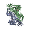
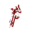
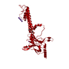
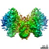
| Title | Cryo-EM structure of the G protein-gated inward rectifier K+ channel GIRK2 (Kir3.2) in complex with PIP2 | ||||||
 Components Components | G protein-activated inward rectifier potassium channel 2 | ||||||
 Keywords Keywords | MEMBRANE PROTEIN / G protein-coupled inwardly rectifying potassium channels / PIP2 | ||||||
| Function / homology |  Function and homology information Function and homology informationG-protein activated inward rectifier potassium channel activity / Activation of G protein gated Potassium channels / Inhibition of voltage gated Ca2+ channels via Gbeta/gamma subunits / inward rectifier potassium channel complex / voltage-gated monoatomic ion channel activity involved in regulation of presynaptic membrane potential / inward rectifier potassium channel activity / regulation of monoatomic ion transmembrane transport / neuronal cell body membrane / potassium ion import across plasma membrane / parallel fiber to Purkinje cell synapse ...G-protein activated inward rectifier potassium channel activity / Activation of G protein gated Potassium channels / Inhibition of voltage gated Ca2+ channels via Gbeta/gamma subunits / inward rectifier potassium channel complex / voltage-gated monoatomic ion channel activity involved in regulation of presynaptic membrane potential / inward rectifier potassium channel activity / regulation of monoatomic ion transmembrane transport / neuronal cell body membrane / potassium ion import across plasma membrane / parallel fiber to Purkinje cell synapse / G-protein alpha-subunit binding / potassium channel activity / negative regulation of insulin secretion / presynapse / presynaptic membrane / postsynapse / axon / dendrite / cell surface / membrane / plasma membrane Similarity search - Function | ||||||
| Biological species |  | ||||||
| Method | ELECTRON MICROSCOPY / single particle reconstruction / cryo EM / Resolution: 3.3 Å | ||||||
 Authors Authors | Niu, Y. / Tao, X. / MacKinnon, R. | ||||||
| Funding support |  United States, 1items United States, 1items
| ||||||
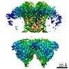
 Citation Citation | Journal: Elife / Year: 2020 Title: Cryo-EM analysis of PIP regulation in mammalian GIRK channels. Authors: Yiming Niu / Xiao Tao / Kouki K Touhara / Roderick MacKinnon / Abstract: G-protein-gated inward rectifier potassium (GIRK) channels are regulated by G proteins and PIP. Here, using cryo-EM single particle analysis we describe the equilibrium ensemble of structures of ...G-protein-gated inward rectifier potassium (GIRK) channels are regulated by G proteins and PIP. Here, using cryo-EM single particle analysis we describe the equilibrium ensemble of structures of neuronal GIRK2 as a function of the C8-PIP concentration. We find that PIP shifts the equilibrium between two distinguishable structures of neuronal GIRK (GIRK2), extended and docked, towards the docked form. In the docked form the cytoplasmic domain, to which G binds, becomes accessible to the cytoplasmic membrane surface where G resides. Furthermore, PIP binding reshapes the G binding surface on the cytoplasmic domain, preparing it to receive G. We find that cardiac GIRK (GIRK1/4) can also exist in both extended and docked conformations. These findings lead us to conclude that PIP influences GIRK channels in a structurally similar manner to Kir2.2 channels. In Kir2.2 channels, the PIP-induced conformational changes open the pore. In GIRK channels, they prepare the channel for activation by G. | ||||||
| History |
|
- Structure visualization
| Movie |
Movie viewer |
|---|---|
| Structure viewer | Molecule: MolmilJmol/JSmol |
- Downloads & links
-Download
| PDBx/mmCIF format | 6xit.cif.gz | 312.2 KB | Display | PDBx/mmCIF format |
|---|---|---|---|---|
| PDB format | pdb6xit.ent.gz | 237 KB | Display | PDB format |
| PDBx/mmJSON format | 6xit.json.gz | Tree view | PDBx/mmJSON format | |
| Others |  Other downloads Other downloads |
-Validation report
| Arichive directory | https://data.pdbj.org/pub/pdb/validation_reports/xi/6xitftp://data.pdbj.org/pub/pdb/validation_reports/xi/6xit | HTTPS FTP |
|---|
-Related structure data
| Related structure data |  22200MC 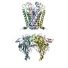 6xisC C: citing same article ( M: map data used to model this data |
|---|---|
| Similar structure data |


-Links
 PDBj
PDBj
- Assembly
| Deposited unit | 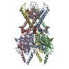
|
|---|---|
| 1 |
|
-Components
| #1: Protein | Mass: 39061.957 Da / Num. of mol.: 4 Source method: isolated from a genetically manipulated source Source: (gene. exp.)  Komagataella pastoris (fungus) / References: UniProt: A0A338P6L0, UniProt: P48542*PLUS Komagataella pastoris (fungus) / References: UniProt: A0A338P6L0, UniProt: P48542*PLUS#2: Chemical | ChemComp-PIO / [(   Mass: 746.566 Da / Num. of mol.: 4 / Source method: obtained synthetically / Formula: C25H49O19P3 / Feature type: SUBJECT OF INVESTIGATION Mass: 746.566 Da / Num. of mol.: 4 / Source method: obtained synthetically / Formula: C25H49O19P3 / Feature type: SUBJECT OF INVESTIGATION#3: Chemical |   Mass: 39.098 Da / Num. of mol.: 3 / Source method: obtained synthetically / Formula: K / Feature type: SUBJECT OF INVESTIGATION Mass: 39.098 Da / Num. of mol.: 3 / Source method: obtained synthetically / Formula: K / Feature type: SUBJECT OF INVESTIGATIONHas ligand of interest | Y | Has protein modification | Y | |
|---|
-Experimental details
-Experiment
| Experiment | Method: ELECTRON MICROSCOPY |
|---|---|
| EM experiment | Aggregation state: PARTICLE / 3D reconstruction method: single particle reconstruction |
- Sample preparation
| Component | Name: Homo-tetrameric assembly of G protein-activated inward rectifier potassium channel 2 Type: COMPLEX / Entity ID: #1 / Source: RECOMBINANT |
|---|---|
| Molecular weight | Experimental value: NO |
| Source (natural) | Organism: |
| Source (recombinant) | Organism: Komagataella pastoris (fungus) |
| Buffer solution | pH: 7.5 Details: 20 mM Tris-HCl pH 7.5, 150 mM KCl, 10 mM DTT, 1 mM DETA, 0.2% DM and 0.01% CHS |
| Specimen | Conc.: 6 mg/ml / Embedding applied: NO / Shadowing applied: NO / Staining applied: NO / Vitrification applied: YES |
| Specimen support | Grid material: GOLD / Grid mesh size: 400 divisions/in. / Grid type: Quantifoil R1.2/1.3 |
| Vitrification | Instrument: FEI VITROBOT MARK IV / Cryogen name: ETHANE / Humidity: 95 % / Chamber temperature: 298 K |
- Electron microscopy imaging
| Experimental equipment |  Model: Titan Krios / Image courtesy: FEI Company |
|---|---|
| Microscopy | Model: FEI TITAN KRIOS |
| Electron gun | Electron source:  FIELD EMISSION GUN / Accelerating voltage: 300 kV / Illumination mode: FLOOD BEAM FIELD EMISSION GUN / Accelerating voltage: 300 kV / Illumination mode: FLOOD BEAM |
| Electron lens | Mode: BRIGHT FIELD / C2 aperture diameter: 100 µm |
| Specimen holder | Cryogen: NITROGEN / Specimen holder model: FEI TITAN KRIOS AUTOGRID HOLDER |
| Image recording | Average exposure time: 10 sec. / Electron dose: 8 e/Å2 / Detector mode: SUPER-RESOLUTION / Film or detector model: GATAN K2 SUMMIT (4k x 4k) / Num. of grids imaged: 1 / Num. of real images: 11349 |
| Image scans | Movie frames/image: 50 / Used frames/image: 1-50 |
- Processing
| EM software |
| ||||||||||||||||||||||||||||||||||||||||||||||||
|---|---|---|---|---|---|---|---|---|---|---|---|---|---|---|---|---|---|---|---|---|---|---|---|---|---|---|---|---|---|---|---|---|---|---|---|---|---|---|---|---|---|---|---|---|---|---|---|---|---|
| CTF correction | Type: NONE | ||||||||||||||||||||||||||||||||||||||||||||||||
| Symmetry | Point symmetry: C4 (4 fold cyclic) | ||||||||||||||||||||||||||||||||||||||||||||||||
| 3D reconstruction | Resolution: 3.3 Å / Resolution method: FSC 0.143 CUT-OFF / Num. of particles: 155128 / Symmetry type: POINT | ||||||||||||||||||||||||||||||||||||||||||||||||
| Atomic model building | PDB-ID: 3SYA Pdb chain-ID: A / Accession code: 3SYA / Pdb chain residue range: 53-380 / Source name: PDB / Type: experimental model |

