 Movie
Movie Controller

[English] 日本語
 Yorodumi
- PDB-6xgy: Crystal structure of E. coli MlaFB ABC transport subunits in the ... -
+ Open data

- Basic information
| Entry | Database: PDB / ID: 6xgy | ||||||||||||||||||
|---|---|---|---|---|---|---|---|---|---|---|---|---|---|---|---|---|---|---|---|
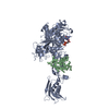
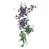
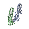
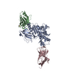
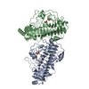
| Title | Crystal structure of E. coli MlaFB ABC transport subunits in the dimeric state | ||||||||||||||||||
 Components Components |
| ||||||||||||||||||
 Keywords Keywords | LIPID TRANSPORT / ABC transporters / MlaF / MlaB / bacterial outer membrane / ATPase / regulation / STAS domain | ||||||||||||||||||
| Function / homology |  Function and homology information Function and homology informationphospholipid transfer activity / intermembrane phospholipid transfer / phospholipid-translocating ATPase complex / phospholipid transport / ATPase-coupled transmembrane transporter activity / response to antibiotic / DNA damage response / ATP binding / membrane / cytoplasm / cytosol Similarity search - Function | ||||||||||||||||||
| Biological species |  | ||||||||||||||||||
| Method |  X-RAY DIFFRACTION / SYNCHROTRON / MOLECULAR REPLACEMENT / Resolution: 2.9 Å X-RAY DIFFRACTION / SYNCHROTRON / MOLECULAR REPLACEMENT / Resolution: 2.9 Å | ||||||||||||||||||
 Authors Authors | Chang, Y. / Bhabha, G. / Ekiert, D.C. | ||||||||||||||||||
| Funding support |  United States, 5items United States, 5items
| ||||||||||||||||||
 Citation Citation | Journal: Elife / Year: 2020 Title: Structure of MlaFB uncovers novel mechanisms of ABC transporter regulation. Authors: Kolich, L.R. / Chang, Y.T. / Coudray, N. / Giacometti, S.I. / MacRae, M.R. / Isom, G.L. / Teran, E.M. / Bhabha, G. / Ekiert, D.C. | ||||||||||||||||||
| History |
|
- Structure visualization
| Structure viewer | Molecule: MolmilJmol/JSmol |
|---|
- Downloads & links
-Download
| PDBx/mmCIF format | 6xgy.cif.gz | 161.7 KB | Display | PDBx/mmCIF format |
|---|---|---|---|---|
| PDB format | pdb6xgy.ent.gz | 125 KB | Display | PDB format |
| PDBx/mmJSON format | 6xgy.json.gz | Tree view | PDBx/mmJSON format | |
| Others |  Other downloads Other downloads |
-Validation report
| Arichive directory | https://data.pdbj.org/pub/pdb/validation_reports/xg/6xgyftp://data.pdbj.org/pub/pdb/validation_reports/xg/6xgy | HTTPS FTP |
|---|
-Related structure data
| Related structure data |  6xgzC  2oukS  3f43S S: Starting model for refinement C: citing same article ( |
|---|---|
| Similar structure data |

-Links
 PDBj
PDBj


- Assembly
| Deposited unit | 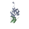
| ||||||||
|---|---|---|---|---|---|---|---|---|---|
| 1 | 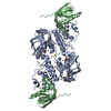
| ||||||||
| Unit cell |
|
-Components
| #1: Protein | Mass: 29128.801 Da / Num. of mol.: 1 Source method: isolated from a genetically manipulated source Source: (gene. exp.) | ||||
|---|---|---|---|---|---|
| #2: Protein | Mass: 12314.044 Da / Num. of mol.: 1 Source method: isolated from a genetically manipulated source Source: (gene. exp.) | ||||
| #3: Chemical | ChemComp-ADP /   Mass: 427.201 Da / Num. of mol.: 1 / Source method: obtained synthetically / Formula: C10H15N5O10P2 / Feature type: SUBJECT OF INVESTIGATION / Comment: ADP, energy-carrying molecule*YM Mass: 427.201 Da / Num. of mol.: 1 / Source method: obtained synthetically / Formula: C10H15N5O10P2 / Feature type: SUBJECT OF INVESTIGATION / Comment: ADP, energy-carrying molecule*YM | ||||
| #4: Chemical |   Mass: 24.305 Da / Num. of mol.: 2 / Source method: obtained synthetically / Formula: Mg Mass: 24.305 Da / Num. of mol.: 2 / Source method: obtained synthetically / Formula: Mg#5: Water | ChemComp-HOH / |  Mass: 18.015 Da / Num. of mol.: 4 / Source method: isolated from a natural source / Formula: H2O Mass: 18.015 Da / Num. of mol.: 4 / Source method: isolated from a natural source / Formula: H2OHas ligand of interest | Y | |
-Experimental details
-Experiment
| Experiment | Method: X-RAY DIFFRACTION / Number of used crystals: 1 |
|---|
- Sample preparation
| Crystal | Density Matthews: 3.3 Å3/Da / Density % sol: 62.68 % |
|---|---|
| Crystal grow | Temperature: 277.15 K / Method: vapor diffusion, sitting drop / Details: 0.2 M magnesium formate |
-Data collection
| Diffraction | Mean temperature: 100 K / Serial crystal experiment: N |
|---|---|
| Diffraction source | Source: SYNCHROTRON / Site: ALS / Beamline: 8.3.1 / Wavelength: 1.11583 Å |
| Detector | Type: DECTRIS PILATUS3 S 6M / Detector: PIXEL / Date: Aug 12, 2016 |
| Radiation | Monochromator: Water-cooled flat double Si(111) Khozu monochromator Protocol: SINGLE WAVELENGTH / Monochromatic (M) / Laue (L): M / Scattering type: x-ray |
| Radiation wavelength | Wavelength: 1.11583 Å / Relative weight: 1 |
| Reflection | Resolution: 2.9→44.57 Å / Num. obs: 12466 / % possible obs: 93.09 % / Redundancy: 19.7 % / CC1/2: 0.999 / Net I/σ(I): 16.09 |
| Reflection shell | Resolution: 2.9→3.004 Å / Num. unique obs: 1213 / CC1/2: 0.349 |
- Processing
| Software |
| ||||||||||||||||||||||||||||||||||||||||
|---|---|---|---|---|---|---|---|---|---|---|---|---|---|---|---|---|---|---|---|---|---|---|---|---|---|---|---|---|---|---|---|---|---|---|---|---|---|---|---|---|---|
| Refinement | Method to determine structure: MOLECULAR REPLACEMENT Starting model: 2OUK, 3F43 Resolution: 2.9→44.57 Å / SU ML: 0.39 / Cross valid method: THROUGHOUT / σ(F): 0 / Phase error: 27.37 / Stereochemistry target values: ML
| ||||||||||||||||||||||||||||||||||||||||
| Solvent computation | Shrinkage radii: 1 Å / VDW probe radii: 1.3 Å / Solvent model: FLAT BULK SOLVENT MODEL | ||||||||||||||||||||||||||||||||||||||||
| Displacement parameters | Biso max: 181.18 Å2 / Biso mean: 84.1482 Å2 / Biso min: 36.55 Å2 | ||||||||||||||||||||||||||||||||||||||||
| Refinement step | Cycle: final / Resolution: 2.9→44.57 Å
| ||||||||||||||||||||||||||||||||||||||||
| LS refinement shell | Refine-ID: X-RAY DIFFRACTION / Rfactor Rfree error: 0 / Total num. of bins used: 4
| ||||||||||||||||||||||||||||||||||||||||
| Refinement TLS params. | Method: refined / Origin x: -35.9284 Å / Origin y: 17.9383 Å / Origin z: 3.3316 Å
| ||||||||||||||||||||||||||||||||||||||||
| Refinement TLS group |
|
