+ Open data
- Basic information
| Entry | Database: PDB / ID: 6xdc | |||||||||
|---|---|---|---|---|---|---|---|---|---|---|
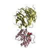
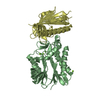
| Title | Cryo-EM structure of SARS-CoV-2 ORF3a | |||||||||
 Components Components | ORF3a protein | |||||||||
 Keywords Keywords | TRANSPORT PROTEIN / SARS-CoV-2 / coronavirus / viroporin / ion channel | |||||||||
| Function / homology |  Function and homology information Function and homology informationhost cell lysosome / symbiont-mediated activation of host reticulophagy / Maturation of protein 3a / SARS-CoV-2 modulates autophagy / : / voltage-gated calcium channel complex / host cell endoplasmic reticulum / monoatomic ion channel activity / SARS-CoV-2 targets host intracellular signalling and regulatory pathways / voltage-gated potassium channel complex ...host cell lysosome / symbiont-mediated activation of host reticulophagy / Maturation of protein 3a / SARS-CoV-2 modulates autophagy / : / voltage-gated calcium channel complex / host cell endoplasmic reticulum / monoatomic ion channel activity / SARS-CoV-2 targets host intracellular signalling and regulatory pathways / voltage-gated potassium channel complex / molecular function activator activity / cytoplasmic side of plasma membrane / host cell endosome / Translation of Structural Proteins / Virion Assembly and Release / Induction of Cell-Cell Fusion / Attachment and Entry / host cell endoplasmic reticulum membrane / SARS-CoV-2 activates/modulates innate and adaptive immune responses / host cell plasma membrane / virion membrane / extracellular region / identical protein binding / plasma membrane Similarity search - Function | |||||||||
| Biological species |  | |||||||||
| Method | ELECTRON MICROSCOPY / single particle reconstruction / cryo EM / Resolution: 2.9 Å | |||||||||
 Authors Authors | Kern, D.M. / Hoel, C.M. / Brohawn, S.G. | |||||||||
| Funding support |  United States, 2items United States, 2items
| |||||||||
 Citation Citation |  Journal: Nat Struct Mol Biol / Year: 2021 Journal: Nat Struct Mol Biol / Year: 2021Title: Cryo-EM structure of SARS-CoV-2 ORF3a in lipid nanodiscs. Authors: David M Kern / Ben Sorum / Sonali S Mali / Christopher M Hoel / Savitha Sridharan / Jonathan P Remis / Daniel B Toso / Abhay Kotecha / Diana M Bautista / Stephen G Brohawn /   Abstract: SARS-CoV-2 ORF3a is a putative viral ion channel implicated in autophagy inhibition, inflammasome activation and apoptosis. 3a protein and anti-3a antibodies are found in infected patient tissues and ...SARS-CoV-2 ORF3a is a putative viral ion channel implicated in autophagy inhibition, inflammasome activation and apoptosis. 3a protein and anti-3a antibodies are found in infected patient tissues and plasma. Deletion of 3a in SARS-CoV-1 reduces viral titer and morbidity in mice, suggesting it could be an effective target for vaccines or therapeutics. Here, we present structures of SARS-CoV-2 3a determined by cryo-EM to 2.1-Å resolution. 3a adopts a new fold with a polar cavity that opens to the cytosol and membrane through separate water- and lipid-filled openings. Hydrophilic grooves along outer helices could form ion-conduction paths. Using electrophysiology and fluorescent ion imaging of 3a-reconstituted liposomes, we observe Ca-permeable, nonselective cation channel activity, identify mutations that alter ion permeability and discover polycationic inhibitors of 3a activity. 3a-like proteins are found across coronavirus lineages that infect bats and humans, suggesting that 3a-targeted approaches could treat COVID-19 and other coronavirus diseases. #1: Journal: bioRxiv / Year: 2021 Title: Cryo-EM structure of the SARS-CoV-2 3a ion channel in lipid nanodiscs. Authors: David M Kern / Ben Sorum / Sonali S Mali / Christopher M Hoel / Savitha Sridharan / Jonathan P Remis / Daniel B Toso / Abhay Kotecha / Diana M Bautista / Stephen G Brohawn /   Abstract: Severe acute respiratory syndrome coronavirus 2 (SARS-CoV-2) is the virus that causes the coronavirus disease 2019 (COVID-19). SARS-CoV-2 encodes three putative ion channels: E, 8a, and 3a. 3a is ...Severe acute respiratory syndrome coronavirus 2 (SARS-CoV-2) is the virus that causes the coronavirus disease 2019 (COVID-19). SARS-CoV-2 encodes three putative ion channels: E, 8a, and 3a. 3a is expressed in SARS patient tissue and anti-3a antibodies are observed in patient plasma. 3a has been implicated in viral release, inhibition of autophagy, inflammasome activation, and cell death and its deletion reduces viral titer and morbidity in mice, raising the possibility that 3a could be an effective vaccine or therapeutic target. Here, we present the first cryo-EM structures of SARS-CoV-2 3a to 2.1 Å resolution and demonstrate 3a forms an ion channel in reconstituted liposomes. The structures in lipid nanodiscs reveal 3a dimers and tetramers adopt a novel fold with a large polar cavity that spans halfway across the membrane and is accessible to the cytosol and the surrounding bilayer through separate water- and lipid-filled openings. Electrophysiology and fluorescent ion imaging experiments show 3a forms Ca-permeable non-selective cation channels. We identify point mutations that alter ion permeability and discover polycationic inhibitors of 3a channel activity. We find 3a-like proteins in multiple and lineages that infect bats and humans. These data show 3a forms a functional ion channel that may promote COVID-19 pathogenesis and suggest targeting 3a could broadly treat coronavirus diseases. | |||||||||
| History |
|
- Structure visualization
| Movie |
 Movie viewer Movie viewer |
|---|---|
| Structure viewer | Molecule:  Molmil Molmil Jmol/JSmol Jmol/JSmol |
- Downloads & links
- Download
| PDBx/mmCIF format |  6xdc.cif.gz 6xdc.cif.gz | 84.1 KB | Display |  PDBx/mmCIF format PDBx/mmCIF format |
|---|---|---|---|---|
| PDB format |  pdb6xdc.ent.gz pdb6xdc.ent.gz | 60.1 KB | Display |  PDB format PDB format |
| PDBx/mmJSON format |  6xdc.json.gz 6xdc.json.gz | Tree view |  PDBx/mmJSON format PDBx/mmJSON format | |
| Others |  Other downloads Other downloads |
-Validation report
| Summary document |  6xdc_validation.pdf.gz 6xdc_validation.pdf.gz | 789.7 KB | Display |  wwPDB validaton report wwPDB validaton report |
|---|---|---|---|---|
| Full document |  6xdc_full_validation.pdf.gz 6xdc_full_validation.pdf.gz | 791.2 KB | Display | |
| Data in XML |  6xdc_validation.xml.gz 6xdc_validation.xml.gz | 17.7 KB | Display | |
| Data in CIF |  6xdc_validation.cif.gz 6xdc_validation.cif.gz | 24.2 KB | Display | |
| Arichive directory |  https://data.pdbj.org/pub/pdb/validation_reports/xd/6xdc https://data.pdbj.org/pub/pdb/validation_reports/xd/6xdc ftp://data.pdbj.org/pub/pdb/validation_reports/xd/6xdc ftp://data.pdbj.org/pub/pdb/validation_reports/xd/6xdc | HTTPS FTP |
-Related structure data
| Related structure data |  22136MC  7kjrC M: map data used to model this data C: citing same article ( |
|---|---|
| Similar structure data | |
| EM raw data |  EMPIAR-10439 (Title: SARS-CoV-2 ORF3a dimer in an MSP1E3D1 lipid nanodisc / Data size: 4.4 TB EMPIAR-10439 (Title: SARS-CoV-2 ORF3a dimer in an MSP1E3D1 lipid nanodisc / Data size: 4.4 TBData #1: Unaligned multi-frame movies of SARS-CoV-2 ORF3a dimer in an MSP1E3D1 lipid nanodisc [micrographs - multiframe]) |
- Links
Links
- Assembly
| Deposited unit | 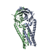
|
|---|---|
| 1 |
|
- Components
| #1: Protein | Mass: 32165.902 Da / Num. of mol.: 2 Source method: isolated from a genetically manipulated source Source: (gene. exp.)  Gene: 3a / Production host:  |
|---|
-Experimental details
-Experiment
| Experiment | Method: ELECTRON MICROSCOPY |
|---|---|
| EM experiment | Aggregation state: PARTICLE / 3D reconstruction method: single particle reconstruction |
- Sample preparation
| Component | Name: ORF3a in MSPE3D1 nanodisc / Type: COMPLEX / Entity ID: all / Source: RECOMBINANT | |||||||||||||||
|---|---|---|---|---|---|---|---|---|---|---|---|---|---|---|---|---|
| Molecular weight | Value: 0.064 MDa / Experimental value: NO | |||||||||||||||
| Source (natural) | Organism:  | |||||||||||||||
| Source (recombinant) | Organism:  | |||||||||||||||
| Buffer solution | pH: 7.4 | |||||||||||||||
| Buffer component |
| |||||||||||||||
| Specimen | Conc.: 1.1 mg/ml / Embedding applied: NO / Shadowing applied: NO / Staining applied: NO / Vitrification applied: YES | |||||||||||||||
| Specimen support | Grid material: GOLD / Grid mesh size: 300 divisions/in. / Grid type: Quantifoil | |||||||||||||||
| Vitrification | Instrument: FEI VITROBOT MARK IV / Cryogen name: ETHANE / Humidity: 100 % / Chamber temperature: 277 K Details: 1 blot force, 5 second wait time, 3 second blot time |
- Electron microscopy imaging
| Experimental equipment |  Model: Talos Arctica / Image courtesy: FEI Company |
|---|---|
| Microscopy | Model: FEI TALOS ARCTICA |
| Electron gun | Electron source:  FIELD EMISSION GUN / Accelerating voltage: 200 kV / Illumination mode: FLOOD BEAM FIELD EMISSION GUN / Accelerating voltage: 200 kV / Illumination mode: FLOOD BEAM |
| Electron lens | Mode: BRIGHT FIELD / Cs: 2.7 mm / C2 aperture diameter: 50 µm / Alignment procedure: COMA FREE |
| Specimen holder | Cryogen: NITROGEN / Specimen holder model: FEI TITAN KRIOS AUTOGRID HOLDER |
| Image recording | Average exposure time: 5 sec. / Electron dose: 50.675 e/Å2 / Film or detector model: GATAN K3 (6k x 4k) / Num. of grids imaged: 1 / Num. of real images: 2595 / Details: 50 frames per image |
| Image scans | Width: 11520 / Height: 8184 |
- Processing
| Software |
| ||||||||||||||||||||||||||||||||||||
|---|---|---|---|---|---|---|---|---|---|---|---|---|---|---|---|---|---|---|---|---|---|---|---|---|---|---|---|---|---|---|---|---|---|---|---|---|---|
| EM software |
| ||||||||||||||||||||||||||||||||||||
| CTF correction | Type: PHASE FLIPPING AND AMPLITUDE CORRECTION | ||||||||||||||||||||||||||||||||||||
| Particle selection | Num. of particles selected: 4134279 | ||||||||||||||||||||||||||||||||||||
| Symmetry | Point symmetry: C2 (2 fold cyclic) | ||||||||||||||||||||||||||||||||||||
| 3D reconstruction | Resolution: 2.9 Å / Resolution method: FSC 0.143 CUT-OFF / Num. of particles: 185871 / Symmetry type: POINT | ||||||||||||||||||||||||||||||||||||
| Atomic model building | B value: 108.61 / Protocol: OTHER / Space: REAL | ||||||||||||||||||||||||||||||||||||
| Refinement | Cross valid method: NONE Stereochemistry target values: GeoStd + Monomer Library + CDL v1.2 | ||||||||||||||||||||||||||||||||||||
| Displacement parameters | Biso mean: 108.61 Å2 | ||||||||||||||||||||||||||||||||||||
| Refine LS restraints |
|
 Movie
Movie Controller







 PDBj
PDBj