 Movie
Movie Controller


+ Open data

- Basic information
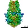
| Entry | Database: EMDB / ID: EMD-22136 | |||||||||
|---|---|---|---|---|---|---|---|---|---|---|
| Title | Cryo-EM structure of SARS-CoV-2 ORF3a | |||||||||
 Map data Map data | SARS-CoV-2 3A | |||||||||
 Sample Sample |
| |||||||||
 Keywords Keywords | SARS-CoV-2 / coronavirus / viroporin / ion channel / TRANSPORT PROTEIN | |||||||||
| Function / homology |  Function and homology information Function and homology informationhost cell lysosome / symbiont-mediated activation of host reticulophagy / Maturation of protein 3a / SARS-CoV-2 modulates autophagy / : / voltage-gated calcium channel complex / host cell endoplasmic reticulum / monoatomic ion channel activity / SARS-CoV-2 targets host intracellular signalling and regulatory pathways / voltage-gated potassium channel complex ...host cell lysosome / symbiont-mediated activation of host reticulophagy / Maturation of protein 3a / SARS-CoV-2 modulates autophagy / : / voltage-gated calcium channel complex / host cell endoplasmic reticulum / monoatomic ion channel activity / SARS-CoV-2 targets host intracellular signalling and regulatory pathways / voltage-gated potassium channel complex / molecular function activator activity / cytoplasmic side of plasma membrane / Translation of Structural Proteins / Virion Assembly and Release / host cell endosome / Induction of Cell-Cell Fusion / Attachment and Entry / host cell endoplasmic reticulum membrane / host cell plasma membrane / SARS-CoV-2 activates/modulates innate and adaptive immune responses / virion membrane / extracellular region / identical protein binding / plasma membrane Similarity search - Function | |||||||||
| Biological species |   Severe acute respiratory syndrome coronavirus 2 Severe acute respiratory syndrome coronavirus 2 | |||||||||
| Method | single particle reconstruction / cryo EM / Resolution: 2.9 Å | |||||||||
 Authors Authors | Kern DM / Hoel CM | |||||||||
| Funding support |  United States, 2 items United States, 2 items
| |||||||||
 Citation Citation | Journal: bioRxiv / Year: 2021 Title: Cryo-EM structure of the SARS-CoV-2 3a ion channel in lipid nanodiscs. Authors: David M Kern / Ben Sorum / Sonali S Mali / Christopher M Hoel / Savitha Sridharan / Jonathan P Remis / Daniel B Toso / Abhay Kotecha / Diana M Bautista / Stephen G Brohawn /  Abstract: Severe acute respiratory syndrome coronavirus 2 (SARS-CoV-2) is the virus that causes the coronavirus disease 2019 (COVID-19). SARS-CoV-2 encodes three putative ion channels: E, 8a, and 3a. 3a is ...Severe acute respiratory syndrome coronavirus 2 (SARS-CoV-2) is the virus that causes the coronavirus disease 2019 (COVID-19). SARS-CoV-2 encodes three putative ion channels: E, 8a, and 3a. 3a is expressed in SARS patient tissue and anti-3a antibodies are observed in patient plasma. 3a has been implicated in viral release, inhibition of autophagy, inflammasome activation, and cell death and its deletion reduces viral titer and morbidity in mice, raising the possibility that 3a could be an effective vaccine or therapeutic target. Here, we present the first cryo-EM structures of SARS-CoV-2 3a to 2.1 Å resolution and demonstrate 3a forms an ion channel in reconstituted liposomes. The structures in lipid nanodiscs reveal 3a dimers and tetramers adopt a novel fold with a large polar cavity that spans halfway across the membrane and is accessible to the cytosol and the surrounding bilayer through separate water- and lipid-filled openings. Electrophysiology and fluorescent ion imaging experiments show 3a forms Ca-permeable non-selective cation channels. We identify point mutations that alter ion permeability and discover polycationic inhibitors of 3a channel activity. We find 3a-like proteins in multiple and lineages that infect bats and humans. These data show 3a forms a functional ion channel that may promote COVID-19 pathogenesis and suggest targeting 3a could broadly treat coronavirus diseases. | |||||||||
| History |
|
- Structure visualization
| Movie |
Movie viewer |
|---|---|
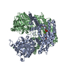
| Structure viewer | EM map: SurfViewMolmilJmol/JSmol |
| Supplemental images |

- Downloads & links
-EMDB archive
| Map data | emd_22136.map.gz | 59.7 MB | EMDB map data format | |
|---|---|---|---|---|
| Header (meta data) | emd-22136-v30.xmlemd-22136.xml | 15.5 KB 15.5 KB | Display Display | EMDB header |
| Images | 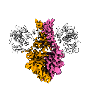 emd_22136.png emd_22136.png | 106.1 KB | ||
| Filedesc metadata | emd-22136.cif.gz | 5.9 KB | ||
| Archive directory |  http://ftp.pdbj.org/pub/emdb/structures/EMD-22136ftp://ftp.pdbj.org/pub/emdb/structures/EMD-22136 http://ftp.pdbj.org/pub/emdb/structures/EMD-22136ftp://ftp.pdbj.org/pub/emdb/structures/EMD-22136 | HTTPS FTP |
-Related structure data
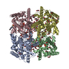
| Related structure data |  6xdcMC  7kjrC C: citing same article ( M: atomic model generated by this map |
|---|---|
| Similar structure data | |
| EM raw data | EMPIAR-10439 (Title: SARS-CoV-2 ORF3a dimer in an MSP1E3D1 lipid nanodisc / Data size: 4.4 TB Data #1: Unaligned multi-frame movies of SARS-CoV-2 ORF3a dimer in an MSP1E3D1 lipid nanodisc [micrographs - multiframe]) |


-Links
| EMDB pages | EMDB (EBI/PDBe) / EMDataResource |
|---|
-Map
| File | Download / File: emd_22136.map.gz / Format: CCP4 / Size: 64 MB / Type: IMAGE STORED AS FLOATING POINT NUMBER (4 BYTES) | ||||||||||||||||||||||||||||||||||||||||||||||||||||||||||||||||||||
|---|---|---|---|---|---|---|---|---|---|---|---|---|---|---|---|---|---|---|---|---|---|---|---|---|---|---|---|---|---|---|---|---|---|---|---|---|---|---|---|---|---|---|---|---|---|---|---|---|---|---|---|---|---|---|---|---|---|---|---|---|---|---|---|---|---|---|---|---|---|
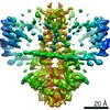
| Annotation | SARS-CoV-2 3A | ||||||||||||||||||||||||||||||||||||||||||||||||||||||||||||||||||||
| Projections & slices | Image control
Images are generated by Spider. | ||||||||||||||||||||||||||||||||||||||||||||||||||||||||||||||||||||
| Voxel size | X=Y=Z: 1.137 Å | ||||||||||||||||||||||||||||||||||||||||||||||||||||||||||||||||||||
| Density |
| ||||||||||||||||||||||||||||||||||||||||||||||||||||||||||||||||||||
| Symmetry | Space group: 1 | ||||||||||||||||||||||||||||||||||||||||||||||||||||||||||||||||||||
| Details | EMDB XML:
CCP4 map header:
| ||||||||||||||||||||||||||||||||||||||||||||||||||||||||||||||||||||
 Z (Sec.)
 Y (Row.)
 X (Col.)


-Supplemental data
- Sample components
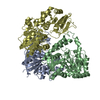
-Entire : ORF3a in MSPE3D1 nanodisc
| Entire | Name: ORF3a in MSPE3D1 nanodisc |
|---|---|
| Components |
|
-Supramolecule #1: ORF3a in MSPE3D1 nanodisc
| Supramolecule | Name: ORF3a in MSPE3D1 nanodisc / type: complex / ID: 1 / Parent: 0 / Macromolecule list: all |
|---|---|
| Source (natural) | Organism: Severe acute respiratory syndrome coronavirus 2 |
| Molecular weight | Theoretical: 64 KDa |
-Macromolecule #1: ORF3a protein
| Macromolecule | Name: ORF3a protein / type: protein_or_peptide / ID: 1 / Number of copies: 2 / Enantiomer: LEVO |
|---|---|
| Source (natural) | Organism: Severe acute respiratory syndrome coronavirus 2 |
| Molecular weight | Theoretical: 32.165902 KDa |
| Recombinant expression | Organism:   Spodoptera frugiperda (fall armyworm) Spodoptera frugiperda (fall armyworm) |
| Sequence | String: MDLFMRIFTI GTVTLKQGEI KDATPSDFVR ATATIPIQAS LPFGWLIVGV ALLAVFQSAS KIITLKKRWQ LALSKGVHFV CNLLLLFVT VYSHLLLVAA GLEAPFLYLY ALVYFLQSIN FVRIIMRLWL CWKCRSKNPL LYDANYFLCW HTNCYDYCIP Y NSVTSSIV ...String: MDLFMRIFTI GTVTLKQGEI KDATPSDFVR ATATIPIQAS LPFGWLIVGV ALLAVFQSAS KIITLKKRWQ LALSKGVHFV CNLLLLFVT VYSHLLLVAA GLEAPFLYLY ALVYFLQSIN FVRIIMRLWL CWKCRSKNPL LYDANYFLCW HTNCYDYCIP Y NSVTSSIV ITSGDGTTSP ISEHDYQIGG YTEKWESGVK DCVVLHSYFT SDYYQLYSTQ LSTDTGVEHV TFFIYNKIVD EP EEHVQIH TIDGSSGVVN PVMEPIYDEP TTTTSVPLSN SLEVLFQ UniProtKB: ORF3a protein |
-Experimental details
-Structure determination
| Method | cryo EM |
|---|---|
 Processing Processing | single particle reconstruction |
| Aggregation state | particle |
-Sample preparation
| Concentration | 1.1 mg/mL | |||||||||
|---|---|---|---|---|---|---|---|---|---|---|
| Buffer | pH: 7.4 Component:
| |||||||||
| Grid | Model: Quantifoil / Material: GOLD / Mesh: 300 / Support film - Material: CARBON / Support film - topology: HOLEY / Support film - Film thickness: 120 / Pretreatment - Type: GLOW DISCHARGE | |||||||||
| Vitrification | Cryogen name: ETHANE / Chamber humidity: 100 % / Chamber temperature: 277 K / Instrument: FEI VITROBOT MARK IV Details: 1 blot force, 5 second wait time, 3 second blot time. |
- Electron microscopy
| Microscope | FEI TALOS ARCTICA |
|---|---|
| Image recording | Film or detector model: GATAN K3 (6k x 4k) / Digitization - Dimensions - Width: 11520 pixel / Digitization - Dimensions - Height: 8184 pixel / Number grids imaged: 1 / Number real images: 2595 / Average exposure time: 5.0 sec. / Average electron dose: 50.675 e/Å2 / Details: 50 frames per image |
| Electron beam | Acceleration voltage: 200 kV / Electron source:  FIELD EMISSION GUN FIELD EMISSION GUN |
| Electron optics | C2 aperture diameter: 50.0 µm / Illumination mode: FLOOD BEAM / Imaging mode: BRIGHT FIELD / Cs: 2.7 mm |
| Sample stage | Specimen holder model: FEI TITAN KRIOS AUTOGRID HOLDER / Cooling holder cryogen: NITROGEN |
| Experimental equipment |  Model: Talos Arctica / Image courtesy: FEI Company |
+Image processing
-Atomic model buiding 1
| Refinement | Space: REAL / Protocol: OTHER / Overall B value: 108.61 |
|---|---|
| Output model | PDB-6xdc: |
