 Movie
Movie Controller

[English] 日本語
 Yorodumi
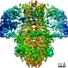
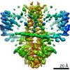
- PDB-6iva: Crystal structure of the S. typhimurium oxaloacetate decarboxylas... -
+ Open data

- Basic information
| Entry | Database: PDB / ID: 6iva | ||||||
|---|---|---|---|---|---|---|---|
| Title | Crystal structure of the S. typhimurium oxaloacetate decarboxylase beta-gamma sub-complex | ||||||
 Components Components |
| ||||||
 Keywords Keywords | MEMBRANE PROTEIN / decarboxylase sodium pump / biotin-dependent decarboxylase | ||||||
| Function / homology |  Function and homology information Function and homology informationoxaloacetate decarboxylase (Na+ extruding) / decarboxylation-driven active transmembrane transporter activity / sodium ion transmembrane transporter activity / oxaloacetate decarboxylase activity / sodium ion export across plasma membrane / sodium ion transport / lyase activity / plasma membrane Similarity search - Function | ||||||
| Biological species |  Salmonella enterica I (bacteria) Salmonella enterica I (bacteria) | ||||||
| Method |  X-RAY DIFFRACTION / SYNCHROTRON / MOLECULAR REPLACEMENT / Resolution: 4.403 Å X-RAY DIFFRACTION / SYNCHROTRON / MOLECULAR REPLACEMENT / Resolution: 4.403 Å | ||||||
 Authors Authors | Xu, X. / Xiang, S. | ||||||
| Funding support |  China, 1items China, 1items
| ||||||
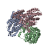
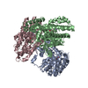
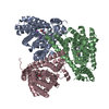
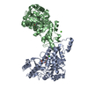
 Citation Citation | Journal: Elife / Year: 2020 Title: Structural insights into sodium transport by the oxaloacetate decarboxylase sodium pump. Authors: Xin Xu / Huigang Shi / Xiaowen Gong / Pu Chen / Ying Gao / Xinzheng Zhang / Song Xiang / Abstract: The oxaloacetate decarboxylase sodium pump (OAD) is a unique primary-active transporter that utilizes the free energy derived from oxaloacetate decarboxylation for sodium transport across the cell ...The oxaloacetate decarboxylase sodium pump (OAD) is a unique primary-active transporter that utilizes the free energy derived from oxaloacetate decarboxylation for sodium transport across the cell membrane. It is composed of 3 subunits: the α subunit catalyzes carboxyl-transfer from oxaloacetate to biotin, the membrane integrated β subunit catalyzes the subsequent carboxyl-biotin decarboxylation and the coupled sodium transport, the γ subunit interacts with the α and β subunits and stabilizes the OAD complex. We present here structure of the OAD βγ sub-complex. The structure revealed that the β and γ subunits form a βγ hetero-hexamer with extensive interactions between the subunits and shed light on the OAD holo-enzyme assembly. Structure-guided functional studies provided insights into the sodium binding sites in the β subunit and the coupling between carboxyl-biotin decarboxylation and sodium transport by the OAD β subunit. | ||||||
| History |
|
- Structure visualization
| Structure viewer | Molecule: MolmilJmol/JSmol |
|---|
- Downloads & links
-Download
| PDBx/mmCIF format | 6iva.cif.gz | 525.5 KB | Display | PDBx/mmCIF format |
|---|---|---|---|---|
| PDB format | pdb6iva.ent.gz | 443.2 KB | Display | PDB format |
| PDBx/mmJSON format | 6iva.json.gz | Tree view | PDBx/mmJSON format | |
| Others |  Other downloads Other downloads |
-Validation report
| Arichive directory | https://data.pdbj.org/pub/pdb/validation_reports/iv/6ivaftp://data.pdbj.org/pub/pdb/validation_reports/iv/6iva | HTTPS FTP |
|---|
-Related structure data
| Related structure data | 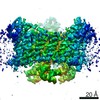 9743C 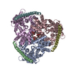 6iwwSC S: Starting model for refinement C: citing same article ( |
|---|---|
| Similar structure data |


-Links
 PDBj
PDBj- Assembly
| Deposited unit | 
| ||||||||
|---|---|---|---|---|---|---|---|---|---|
| 1 |
| ||||||||
| Unit cell |
|
-Components
| #1: Protein | Mass: 44928.801 Da / Num. of mol.: 3 Source method: isolated from a genetically manipulated source Source: (gene. exp.) Salmonella enterica I (bacteria) / Production host: References: UniProt: A0A0F7JAV8, UniProt: Q8ZRY4*PLUS, oxaloacetate decarboxylase (Na+ extruding) #2: Protein | Mass: 11137.029 Da / Num. of mol.: 3 Source method: isolated from a genetically manipulated source Source: (gene. exp.) Salmonella enterica I (bacteria) / Production host: References: UniProt: A0A0F7JC72, UniProt: Q03032*PLUS, oxaloacetate decarboxylase (Na+ extruding) Sequence details | Authors state that this confluence may be a natural occurring mutation that happen to exist in the ...Authors state that this confluence may be a natural occurring mutation that happen to exist in the genomic DNA they used. | |
|---|
-Experimental details
-Experiment
| Experiment | Method: X-RAY DIFFRACTION / Number of used crystals: 1 |
|---|
- Sample preparation
| Crystal | Density Matthews: 3.82 Å3/Da / Density % sol: 67.84 % |
|---|---|
| Crystal grow | Temperature: 295 K / Method: vapor diffusion, sitting drop / pH: 5.5 Details: 0.1 M sodium chloride, 0.1 M lithium sulfate, 0.1 M sodium citrate pH 5.5, 25% PEG1000 and 0.125% PEG400 |
-Data collection
| Diffraction | Mean temperature: 100 K / Serial crystal experiment: N |
|---|---|
| Diffraction source | Source: SYNCHROTRON / Site: SSRF / Beamline: BL17U1 / Wavelength: 0.979 Å |
| Detector | Type: ADSC QUANTUM 315r / Detector: CCD / Date: Jul 12, 2016 |
| Radiation | Protocol: SINGLE WAVELENGTH / Monochromatic (M) / Laue (L): M / Scattering type: x-ray |
| Radiation wavelength | Wavelength: 0.979 Å / Relative weight: 1 |
| Reflection | Resolution: 4.4→50 Å / Num. obs: 16715 / % possible obs: 99.8 % / Redundancy: 6.9 % / CC1/2: 1 / Rmerge(I) obs: 0.16 / Net I/σ(I): 14.99 |
| Reflection shell | Resolution: 4.4→4.48 Å / Mean I/σ(I) obs: 1.52 / Num. unique obs: 834 / CC1/2: 0.549 |
- Processing
| Software |
| |||||||||||||||||||||||||||||||||||||||||||||||||||||||||||||||||||||||||||||||||||||||||||||||||||||||||||||||||||||||||||||||||||||||||||||||||||||||||||||||||||||||||||||||
|---|---|---|---|---|---|---|---|---|---|---|---|---|---|---|---|---|---|---|---|---|---|---|---|---|---|---|---|---|---|---|---|---|---|---|---|---|---|---|---|---|---|---|---|---|---|---|---|---|---|---|---|---|---|---|---|---|---|---|---|---|---|---|---|---|---|---|---|---|---|---|---|---|---|---|---|---|---|---|---|---|---|---|---|---|---|---|---|---|---|---|---|---|---|---|---|---|---|---|---|---|---|---|---|---|---|---|---|---|---|---|---|---|---|---|---|---|---|---|---|---|---|---|---|---|---|---|---|---|---|---|---|---|---|---|---|---|---|---|---|---|---|---|---|---|---|---|---|---|---|---|---|---|---|---|---|---|---|---|---|---|---|---|---|---|---|---|---|---|---|---|---|---|---|---|---|---|
| Refinement | Method to determine structure: MOLECULAR REPLACEMENT Starting model: 6IWW Resolution: 4.403→42.191 Å / SU ML: 0.74 / Cross valid method: FREE R-VALUE / σ(F): 1.33 / Phase error: 45.14
| |||||||||||||||||||||||||||||||||||||||||||||||||||||||||||||||||||||||||||||||||||||||||||||||||||||||||||||||||||||||||||||||||||||||||||||||||||||||||||||||||||||||||||||||
| Solvent computation | Shrinkage radii: 0.9 Å / VDW probe radii: 1.11 Å | |||||||||||||||||||||||||||||||||||||||||||||||||||||||||||||||||||||||||||||||||||||||||||||||||||||||||||||||||||||||||||||||||||||||||||||||||||||||||||||||||||||||||||||||
| Refinement step | Cycle: LAST / Resolution: 4.403→42.191 Å
| |||||||||||||||||||||||||||||||||||||||||||||||||||||||||||||||||||||||||||||||||||||||||||||||||||||||||||||||||||||||||||||||||||||||||||||||||||||||||||||||||||||||||||||||
| Refine LS restraints |
| |||||||||||||||||||||||||||||||||||||||||||||||||||||||||||||||||||||||||||||||||||||||||||||||||||||||||||||||||||||||||||||||||||||||||||||||||||||||||||||||||||||||||||||||
| LS refinement shell |
| |||||||||||||||||||||||||||||||||||||||||||||||||||||||||||||||||||||||||||||||||||||||||||||||||||||||||||||||||||||||||||||||||||||||||||||||||||||||||||||||||||||||||||||||
| Refinement TLS params. | Method: refined / Refine-ID: X-RAY DIFFRACTION
| |||||||||||||||||||||||||||||||||||||||||||||||||||||||||||||||||||||||||||||||||||||||||||||||||||||||||||||||||||||||||||||||||||||||||||||||||||||||||||||||||||||||||||||||
| Refinement TLS group |
|
