Movie
Movie Controller

[English] 日本語
 Yorodumi
- PDB-6wxl: Cryo-EM structure of the VRC315 clinical trial, vaccine-elicited,... -
+ Open data

- Basic information
| Entry | Database: PDB / ID: 6wxl | ||||||
|---|---|---|---|---|---|---|---|
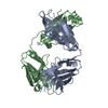
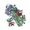
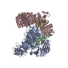
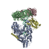
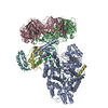
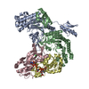
| Title | Cryo-EM structure of the VRC315 clinical trial, vaccine-elicited, human antibody 1D12 in complex with an H7 SH13 HA trimer | ||||||
 Components Components |
| ||||||
 Keywords Keywords | IMMUNE SYSTEM / VRC / VRC315 / Group 2 / Fab | ||||||
| Function / homology |  Function and homology information Function and homology informationviral budding from plasma membrane / clathrin-dependent endocytosis of virus by host cell / host cell surface receptor binding / fusion of virus membrane with host plasma membrane / fusion of virus membrane with host endosome membrane / viral envelope / virion attachment to host cell / host cell plasma membrane / virion membrane / membrane Similarity search - Function | ||||||
| Biological species |   Influenza A virus Influenza A virus Homo sapiens (human) Homo sapiens (human) | ||||||
| Method | ELECTRON MICROSCOPY / single particle reconstruction / cryo EM / Resolution: 2.76 Å | ||||||
 Authors Authors | Gorman, J. / Kwong, P.D. | ||||||
| Funding support |  United States, 1items United States, 1items
| ||||||
 Citation Citation | Journal: Structure / Year: 2022 Title: Structure of an influenza group 2-neutralizing antibody targeting the hemagglutinin stem supersite. Authors: Crystal Sao-Fong Cheung / Jason Gorman / Sarah F Andrews / Reda Rawi / Mateo Reveiz / Chen-Hsiang Shen / Yiran Wang / Darcy R Harris / Alexandra F Nazzari / Adam S Olia / Julie Raab / I-Ting ...Authors: Crystal Sao-Fong Cheung / Jason Gorman / Sarah F Andrews / Reda Rawi / Mateo Reveiz / Chen-Hsiang Shen / Yiran Wang / Darcy R Harris / Alexandra F Nazzari / Adam S Olia / Julie Raab / I-Ting Teng / Raffaello Verardi / Shuishu Wang / Yongping Yang / Gwo-Yu Chuang / Adrian B McDermott / Tongqing Zhou / Peter D Kwong / Abstract: Several influenza antibodies with broad group 2 neutralization have recently been isolated. Here, we analyze the structure, class, and binding of one of these antibodies from an H7N9 vaccine trial, ...Several influenza antibodies with broad group 2 neutralization have recently been isolated. Here, we analyze the structure, class, and binding of one of these antibodies from an H7N9 vaccine trial, 315-19-1D12. The cryo-EM structure of 315-19-1D12 Fab in complex with the hemagglutinin (HA) trimer revealed the antibody to recognize the helix A region of the HA stem, at the supersite of vulnerability recognized by group 1-specific and by cross-group-neutralizing antibodies. 315-19-1D12 was derived from HV1-2 and KV2-28 genes and appeared to form a new antibody class. Bioinformatic analysis indicated its group 2 neutralization specificity to be a consequence of four key residue positions. We specifically tested the impact of the group 1-specific N33 glycan, which decreased but did not abolish group 2 binding of 315-19-1D12. Overall, this study highlights the recognition of a broad group 2-neutralizing antibody, revealing unexpected diversity in neutralization specificity for antibodies that recognize the HA stem supersite. | ||||||
| History |
|
- Structure visualization
| Movie |
Movie viewer |
|---|---|
| Structure viewer | Molecule: MolmilJmol/JSmol |
- Downloads & links
-Download
| PDBx/mmCIF format | 6wxl.cif.gz | 388.3 KB | Display | PDBx/mmCIF format |
|---|---|---|---|---|
| PDB format | pdb6wxl.ent.gz | 311.4 KB | Display | PDB format |
| PDBx/mmJSON format | 6wxl.json.gz | Tree view | PDBx/mmJSON format | |
| Others |  Other downloads Other downloads |
-Validation report
| Arichive directory | https://data.pdbj.org/pub/pdb/validation_reports/wx/6wxlftp://data.pdbj.org/pub/pdb/validation_reports/wx/6wxl | HTTPS FTP |
|---|
-Related structure data
| Related structure data |  21961MC M: map data used to model this data C: citing same article ( |
|---|---|
| Similar structure data |


-Links
 PDBj
PDBj


- Assembly
| Deposited unit | 
|
|---|---|
| 1 |
|
-Components
| #1: Protein | Mass: 35039.473 Da / Num. of mol.: 3 Source method: isolated from a genetically manipulated source Source: (gene. exp.) Influenza A virus (A/Shanghai/JS01/2013(H7N9))Strain: A/Shanghai/JS01/2013(H7N9) / Gene: HA / Production host: Homo sapiens (human) / References: UniProt: A0A067Y6L0#2: Protein | Mass: 25234.352 Da / Num. of mol.: 3 Source method: isolated from a genetically manipulated source Source: (gene. exp.) Influenza A virus (A/Shanghai/JS01/2013(H7N9))Strain: A/Shanghai/JS01/2013(H7N9) / Gene: HA / Production host: Homo sapiens (human) / References: UniProt: A0A067Y6L0#3: Antibody | Mass: 23971.762 Da / Num. of mol.: 3 Source method: isolated from a genetically manipulated source Source: (gene. exp.) Homo sapiens (human) / Production host: Homo sapiens (human)#4: Antibody | Mass: 25042.229 Da / Num. of mol.: 3 Source method: isolated from a genetically manipulated source Source: (gene. exp.) Homo sapiens (human) / Production host: Homo sapiens (human)#5: Sugar | ChemComp-NAG /   Type: D-saccharide, beta linking / Mass: 221.208 Da / Num. of mol.: 9 Type: D-saccharide, beta linking / Mass: 221.208 Da / Num. of mol.: 9Source method: isolated from a genetically manipulated source Formula: C8H15NO6 Has ligand of interest | N | Has protein modification | Y | |
|---|
-Experimental details
-Experiment
| Experiment | Method: ELECTRON MICROSCOPY |
|---|---|
| EM experiment | Aggregation state: PARTICLE / 3D reconstruction method: single particle reconstruction |
- Sample preparation
| Component |
| ||||||||||||||||||||||||
|---|---|---|---|---|---|---|---|---|---|---|---|---|---|---|---|---|---|---|---|---|---|---|---|---|---|
| Molecular weight | Experimental value: NO | ||||||||||||||||||||||||
| Source (natural) |
| ||||||||||||||||||||||||
| Source (recombinant) |
| ||||||||||||||||||||||||
| Buffer solution | pH: 7.4 | ||||||||||||||||||||||||
| Buffer component | Formula: PBS | ||||||||||||||||||||||||
| Specimen | Conc.: 2 mg/ml / Embedding applied: NO / Shadowing applied: NO / Staining applied: NO / Vitrification applied: YES Details: Cryo-EM structure of the VRC315 clinical trial, vaccine-elicited, human antibody 1D12 in complex with an H7 SH13 HA trimer | ||||||||||||||||||||||||
| Specimen support | Grid material: COPPER / Grid type: C-flat-1.2/1.3 4C | ||||||||||||||||||||||||
| Vitrification | Instrument: FEI VITROBOT MARK IV / Cryogen name: ETHANE / Humidity: 90 % / Chamber temperature: 293 K |
- Electron microscopy imaging
| Experimental equipment |  Model: Titan Krios / Image courtesy: FEI Company |
|---|---|
| Microscopy | Model: FEI TITAN KRIOS |
| Electron gun | Electron source:  FIELD EMISSION GUN / Accelerating voltage: 300 kV / Illumination mode: FLOOD BEAM FIELD EMISSION GUN / Accelerating voltage: 300 kV / Illumination mode: FLOOD BEAM |
| Electron lens | Mode: BRIGHT FIELD / C2 aperture diameter: 70 µm |
| Specimen holder | Cryogen: NITROGEN / Specimen holder model: FEI TITAN KRIOS AUTOGRID HOLDER |
| Image recording | Average exposure time: 2 sec. / Electron dose: 49.9 e/Å2 / Detector mode: COUNTING / Film or detector model: GATAN K3 (6k x 4k) / Num. of grids imaged: 1 / Num. of real images: 1878 |
| Image scans | Movie frames/image: 50 |
- Processing
| Software | Name: PHENIX / Version: 1.16_3549: / Classification: refinement | ||||||||||||||||||||||||
|---|---|---|---|---|---|---|---|---|---|---|---|---|---|---|---|---|---|---|---|---|---|---|---|---|---|
| EM software |
| ||||||||||||||||||||||||
| CTF correction | Type: PHASE FLIPPING AND AMPLITUDE CORRECTION | ||||||||||||||||||||||||
| Symmetry | Point symmetry: C3 (3 fold cyclic) | ||||||||||||||||||||||||
| 3D reconstruction | Resolution: 2.76 Å / Resolution method: FSC 0.143 CUT-OFF / Num. of particles: 142628 / Symmetry type: POINT | ||||||||||||||||||||||||
| Atomic model building | Protocol: FLEXIBLE FIT / Space: REAL | ||||||||||||||||||||||||
| Atomic model building |
| ||||||||||||||||||||||||
| Refine LS restraints |
|