 Movie
Movie Controller

[English] 日本語
 Yorodumi
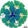
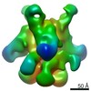
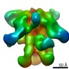
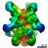
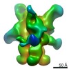
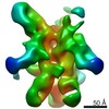
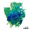
- PDB-6wxe: Cryo-EM reconstruction of VP5*/VP8* assembly from rhesus rotaviru... -
+ Open data

- Basic information
| Entry | Database: PDB / ID: 6wxe | ||||||
|---|---|---|---|---|---|---|---|
| Title | Cryo-EM reconstruction of VP5*/VP8* assembly from rhesus rotavirus particles - Upright conformation | ||||||
 Components Components |
| ||||||
 Keywords Keywords | VIRAL PROTEIN / Complex / non-enveloped virus / viral particle / entry / membrane-penetration / rotavirus / VP4 / VP5* / VP8* | ||||||
| Function / homology |  Function and homology information Function and homology informationviral intermediate capsid / host cell endoplasmic reticulum lumen / host cell rough endoplasmic reticulum / permeabilization of host organelle membrane involved in viral entry into host cell / T=13 icosahedral viral capsid / host cytoskeleton / viral outer capsid / host cell endoplasmic reticulum-Golgi intermediate compartment / receptor-mediated virion attachment to host cell / host cell surface receptor binding ...viral intermediate capsid / host cell endoplasmic reticulum lumen / host cell rough endoplasmic reticulum / permeabilization of host organelle membrane involved in viral entry into host cell / T=13 icosahedral viral capsid / host cytoskeleton / viral outer capsid / host cell endoplasmic reticulum-Golgi intermediate compartment / receptor-mediated virion attachment to host cell / host cell surface receptor binding / fusion of virus membrane with host plasma membrane / viral envelope / virion attachment to host cell / host cell plasma membrane / structural molecule activity / metal ion binding Similarity search - Function | ||||||
| Biological species |  Rotavirus A Rotavirus A | ||||||
| Method | ELECTRON MICROSCOPY / single particle reconstruction / cryo EM / Resolution: 3.4 Å | ||||||
 Authors Authors | Herrmann, T. / Harrison, S.C. / Jenni, S. | ||||||
| Funding support |  United States, 1items United States, 1items
| ||||||
 Citation Citation | Journal: Nature / Year: 2021 Title: Functional refolding of the penetration protein on a non-enveloped virus. Authors: Tobias Herrmann / Raúl Torres / Eric N Salgado / Cristina Berciu / Daniel Stoddard / Daniela Nicastro / Simon Jenni / Stephen C Harrison / Abstract: A non-enveloped virus requires a membrane lesion to deliver its genome into a target cell. For rotaviruses, membrane perforation is a principal function of the viral outer-layer protein, VP4. Here we ...A non-enveloped virus requires a membrane lesion to deliver its genome into a target cell. For rotaviruses, membrane perforation is a principal function of the viral outer-layer protein, VP4. Here we describe the use of electron cryomicroscopy to determine how VP4 performs this function and show that when activated by cleavage to VP8* and VP5*, VP4 can rearrange on the virion surface from an 'upright' to a 'reversed' conformation. The reversed structure projects a previously buried 'foot' domain outwards into the membrane of the host cell to which the virion has attached. Electron cryotomograms of virus particles entering cells are consistent with this picture. Using a disulfide mutant of VP4, we have also stabilized a probable intermediate in the transition between the two conformations. Our results define molecular mechanisms for the first steps of the penetration of rotaviruses into the membranes of target cells and suggest similarities with mechanisms postulated for other viruses. | ||||||
| History |
|
- Structure visualization
| Movie |
Movie viewer |
|---|---|
| Structure viewer | Molecule: MolmilJmol/JSmol |
- Downloads & links
-Download
| PDBx/mmCIF format | 6wxe.cif.gz | 4.4 MB | Display | PDBx/mmCIF format |
|---|---|---|---|---|
| PDB format | pdb6wxe.ent.gz | Display | PDB format | |
| PDBx/mmJSON format | 6wxe.json.gz | Tree view | PDBx/mmJSON format | |
| Others |  Other downloads Other downloads |
-Validation report
| Arichive directory | https://data.pdbj.org/pub/pdb/validation_reports/wx/6wxeftp://data.pdbj.org/pub/pdb/validation_reports/wx/6wxe | HTTPS FTP |
|---|
-Related structure data
| Related structure data |  21955MC  6wxfC  6wxgC M: map data used to model this data C: citing same article ( |
|---|---|
| Similar structure data |

-Links
 PDBj
PDBj


- Assembly
| Deposited unit | 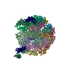
|
|---|---|
| 1 |
|
-Components
| #1: Protein | Mass: 86655.586 Da / Num. of mol.: 3 Source method: isolated from a genetically manipulated source Source: (gene. exp.) Rotavirus A (strain RVA/Monkey/United States/RRV/1975/G3P5B[3])Strain: RVA/Monkey/United States/RRV/1975/G3P5B[3] / Cell line (production host): Sf9 / Production host:   Spodoptera frugiperda (fall armyworm) / References: UniProt: G0YZG6 Spodoptera frugiperda (fall armyworm) / References: UniProt: G0YZG6#2: Protein | Mass: 44934.766 Da / Num. of mol.: 18 / Source method: isolated from a natural source Source: (natural) Rotavirus A (strain RVA/Monkey/United States/RRV/1975/G3P5B[3])Strain: RVA/Monkey/United States/RRV/1975/G3P5B[3] / References: UniProt: B2BN53 #3: Protein | Mass: 37136.531 Da / Num. of mol.: 18 Source method: isolated from a genetically manipulated source Source: (gene. exp.) Rotavirus A (strain RVA/Monkey/United States/RRV/1975/G3P5B[3])Strain: RVA/Monkey/United States/RRV/1975/G3P5B[3] / Cell line (production host): Sf9 / Production host: Spodoptera frugiperda (fall armyworm) / References: UniProt: P12476#4: Sugar | ChemComp-NAG /   Type: D-saccharide, beta linking / Mass: 221.208 Da / Num. of mol.: 18 Type: D-saccharide, beta linking / Mass: 221.208 Da / Num. of mol.: 18Source method: isolated from a genetically manipulated source Formula: C8H15NO6 #5: Chemical | ChemComp-CA /   Mass: 40.078 Da / Num. of mol.: 54 / Source method: obtained synthetically / Formula: Ca Mass: 40.078 Da / Num. of mol.: 54 / Source method: obtained synthetically / Formula: CaHas ligand of interest | N | Has protein modification | Y | |
|---|
-Experimental details
-Experiment
| Experiment | Method: ELECTRON MICROSCOPY |
|---|---|
| EM experiment | Aggregation state: PARTICLE / 3D reconstruction method: single particle reconstruction |
- Sample preparation
| Component | Name: Rotavirus VP4, VP6, VP7 assembly in the spike conformation Type: COMPLEX Details: Obtained from wild-type recoated rhesus rotavirus particles (wt rcTLPs) Entity ID: #1-#3 / Source: MULTIPLE SOURCES | ||||||||||||||||||||
|---|---|---|---|---|---|---|---|---|---|---|---|---|---|---|---|---|---|---|---|---|---|
| Molecular weight | Value: 1.7 MDa / Experimental value: NO | ||||||||||||||||||||
| Source (natural) | Organism: Rotavirus A (strain RVA/Monkey/United States/RRV/1975/G3P5B[3]) | ||||||||||||||||||||
| Source (recombinant) | Organism: Spodoptera frugiperda (fall armyworm) | ||||||||||||||||||||
| Buffer solution | pH: 8 | ||||||||||||||||||||
| Buffer component |
| ||||||||||||||||||||
| Specimen | Conc.: 1.2 mg/ml / Embedding applied: NO / Shadowing applied: NO / Staining applied: NO / Vitrification applied: YES | ||||||||||||||||||||
| Specimen support | Grid material: COPPER / Grid mesh size: 400 divisions/in. / Grid type: C-flat-1.2/1.3 4C | ||||||||||||||||||||
| Vitrification | Instrument: GATAN CRYOPLUNGE 3 / Cryogen name: ETHANE / Humidity: 90 % / Chamber temperature: 298.15 K / Details: 4 ul sample volume, 4 sec blotting time |
- Electron microscopy imaging
| Experimental equipment |  Model: Tecnai Polara / Image courtesy: FEI Company |
|---|---|
| Microscopy | Model: FEI POLARA 300 |
| Electron gun | Electron source:  FIELD EMISSION GUN / Accelerating voltage: 300 kV / Illumination mode: FLOOD BEAM FIELD EMISSION GUN / Accelerating voltage: 300 kV / Illumination mode: FLOOD BEAM |
| Electron lens | Mode: BRIGHT FIELD / Nominal magnification: 27500 X / Calibrated magnification: 40605 X / Nominal defocus max: 3000 nm / Nominal defocus min: 1000 nm / Calibrated defocus min: 1000 nm / Calibrated defocus max: 3000 nm / Cs: 2 mm / Alignment procedure: BASIC |
| Specimen holder | Cryogen: NITROGEN |
| Image recording | Average exposure time: 10 sec. / Electron dose: 33 e/Å2 / Detector mode: COUNTING / Film or detector model: GATAN K2 SUMMIT (4k x 4k) / Num. of grids imaged: 1 / Num. of real images: 4107 |
| Image scans | Width: 3838 / Height: 3701 / Movie frames/image: 50 / Used frames/image: 1-50 |
- Processing
| Software |
| ||||||||||||||||||||||||||||||||||||
|---|---|---|---|---|---|---|---|---|---|---|---|---|---|---|---|---|---|---|---|---|---|---|---|---|---|---|---|---|---|---|---|---|---|---|---|---|---|
| EM software |
| ||||||||||||||||||||||||||||||||||||
| CTF correction | Type: PHASE FLIPPING AND AMPLITUDE CORRECTION | ||||||||||||||||||||||||||||||||||||
| Particle selection | Num. of particles selected: 14579 / Details: whole viral particles | ||||||||||||||||||||||||||||||||||||
| Symmetry | Point symmetry: C1 (asymmetric) | ||||||||||||||||||||||||||||||||||||
| 3D reconstruction | Resolution: 3.4 Å / Resolution method: FSC 0.143 CUT-OFF / Num. of particles: 167147 / Algorithm: FOURIER SPACE Details: Reconstruction final map after classification of VP4 sub-particles. VP4 sub-particles extracted from viral particles (60 VP4 sub-particles per viral particle) Num. of class averages: 1 / Symmetry type: POINT | ||||||||||||||||||||||||||||||||||||
| Atomic model building | Protocol: OTHER / Space: REAL / Details: phenix.real_space_refine | ||||||||||||||||||||||||||||||||||||
| Atomic model building | PDB-ID: 4V7Q Accession code: 4V7Q / Source name: PDB / Type: experimental model | ||||||||||||||||||||||||||||||||||||
| Refinement | Cross valid method: NONE Stereochemistry target values: GeoStd + Monomer Library + CDL v1.2 | ||||||||||||||||||||||||||||||||||||
| Displacement parameters | Biso mean: 26.04 Å2 | ||||||||||||||||||||||||||||||||||||
| Refine LS restraints |
|

