 Movie
Movie Controller


+ Open data

- Basic information
| Entry | Database: PDB / ID: 6ww6 | ||||||
|---|---|---|---|---|---|---|---|
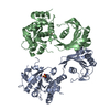
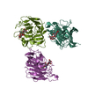
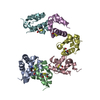
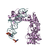
| Title | Crystal structure of EutV bound to RNA | ||||||
 Components Components |
| ||||||
 Keywords Keywords | RNA BINDING PROTEIN/RNA / RNA binding / ANTAR domain / antitermination / RNA BINDING PROTEIN / RNA BINDING PROTEIN-RNA complex | ||||||
| Function / homology |  Function and homology information Function and homology information | ||||||
| Biological species |   Enterococcus faecalis (bacteria) Enterococcus faecalis (bacteria) | ||||||
| Method |  X-RAY DIFFRACTION / SYNCHROTRON / MOLECULAR REPLACEMENT / Resolution: 3.8 Å X-RAY DIFFRACTION / SYNCHROTRON / MOLECULAR REPLACEMENT / Resolution: 3.8 Å | ||||||
 Authors Authors | Ataide, S.F. / Walshe, J.L. | ||||||
 Citation Citation | Journal: Nucleic Acids Res. / Year: 2022 Title: Structural characterization of the ANTAR antiterminator domain bound to RNA. Authors: Walshe, J.L. / Siddiquee, R. / Patel, K. / Ataide, S.F. | ||||||
| History |
|
- Structure visualization
| Structure viewer | Molecule: MolmilJmol/JSmol |
|---|
- Downloads & links
-Download
| PDBx/mmCIF format | 6ww6.cif.gz | 131.7 KB | Display | PDBx/mmCIF format |
|---|---|---|---|---|
| PDB format | pdb6ww6.ent.gz | 96.9 KB | Display | PDB format |
| PDBx/mmJSON format | 6ww6.json.gz | Tree view | PDBx/mmJSON format | |
| Others |  Other downloads Other downloads |
-Validation report
| Arichive directory | https://data.pdbj.org/pub/pdb/validation_reports/ww/6ww6ftp://data.pdbj.org/pub/pdb/validation_reports/ww/6ww6 | HTTPS FTP |
|---|
-Related structure data
| Related structure data | 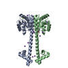 6wshSC S: Starting model for refinement C: citing same article ( |
|---|---|
| Similar structure data |


-Links
 PDBj
PDBj


- Assembly
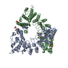
| Deposited unit | 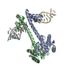
| ||||||||||||
|---|---|---|---|---|---|---|---|---|---|---|---|---|---|
| 1 |
| ||||||||||||
| Unit cell |
|
-Components
| #1: Protein | Mass: 21529.006 Da / Num. of mol.: 2 Source method: isolated from a genetically manipulated source Details: Dimeric EutV bound simultaneously to two seperate dual hexaloops RNA molecules. Each EutV chain binds a single hexaloop from different RNA molecules with the 2nd hexaloop binding to the ...Details: Dimeric EutV bound simultaneously to two seperate dual hexaloops RNA molecules. Each EutV chain binds a single hexaloop from different RNA molecules with the 2nd hexaloop binding to the symmetry related EutV chain in a neighbouring ASUs Source: (gene. exp.) Enterococcus faecalis (bacteria)Gene: BZG32_08120, CUN08_06285, DVW78_01210, FKY84_00940, KUB3007_C13700 Production host: #2: RNA chain | Mass: 17507.568 Da / Num. of mol.: 4 Source method: isolated from a genetically manipulated source Source: (gene. exp.) Enterococcus faecalis (bacteria) / Production host: Cloning vector pUC19 (others) / References: GenBank: 295112306#3: Chemical |   Mass: 66.007 Da / Num. of mol.: 2 / Source method: obtained synthetically / Formula: BeF3 Mass: 66.007 Da / Num. of mol.: 2 / Source method: obtained synthetically / Formula: BeF3Has ligand of interest | N | |
|---|
-Experimental details
-Experiment
| Experiment | Method: X-RAY DIFFRACTION / Number of used crystals: 1 |
|---|
- Sample preparation
| Crystal | Density Matthews: 3.18 Å3/Da / Density % sol: 61.34 % |
|---|---|
| Crystal grow | Temperature: 291.15 K / Method: vapor diffusion, hanging drop / pH: 7.5 Details: 0.1 M Sodium chloride 0.1 M HEPES 1.4-1.8 M Ammonium sulfate |
-Data collection
| Diffraction | Mean temperature: 80 K / Serial crystal experiment: N |
|---|---|
| Diffraction source | Source: SYNCHROTRON / Site: Australian Synchrotron  / Beamline: MX2 / Wavelength: 0.9537 Å / Beamline: MX2 / Wavelength: 0.9537 Å |
| Detector | Type: DECTRIS EIGER X 16M / Detector: PIXEL / Date: Sep 17, 2017 |
| Radiation | Protocol: SINGLE WAVELENGTH / Monochromatic (M) / Laue (L): M / Scattering type: x-ray |
| Radiation wavelength | Wavelength: 0.9537 Å / Relative weight: 1 |
| Reflection | Resolution: 3.8→47.19 Å / Num. obs: 14773 / % possible obs: 99.5 % / Redundancy: 6.7 % / Biso Wilson estimate: 129.7 Å2 / CC1/2: 0.998 / Rmerge(I) obs: 0.151 / Rpim(I) all: 0.084 / Net I/σ(I): 9.1 |
| Reflection shell | Resolution: 3.8→4.25 Å / Redundancy: 6.7 % / Rmerge(I) obs: 0.799 / Mean I/σ(I) obs: 2.3 / Num. unique obs: 4093 / CC1/2: 0.733 / Rpim(I) all: 0.489 / % possible all: 98.9 |
- Processing
| Software |
| ||||||||||||||||||||||||||||||||||||||||||
|---|---|---|---|---|---|---|---|---|---|---|---|---|---|---|---|---|---|---|---|---|---|---|---|---|---|---|---|---|---|---|---|---|---|---|---|---|---|---|---|---|---|---|---|
| Refinement | Method to determine structure: MOLECULAR REPLACEMENT Starting model: 6WSH Resolution: 3.8→47.19 Å / SU ML: 0.4632 / Cross valid method: FREE R-VALUE / σ(F): 1.34 / Phase error: 27.9921 Stereochemistry target values: GeoStd + Monomer Library + CDL v1.2
| ||||||||||||||||||||||||||||||||||||||||||
| Solvent computation | Shrinkage radii: 0.9 Å / VDW probe radii: 1.11 Å / Solvent model: FLAT BULK SOLVENT MODEL | ||||||||||||||||||||||||||||||||||||||||||
| Displacement parameters | Biso mean: 158.4 Å2 | ||||||||||||||||||||||||||||||||||||||||||
| Refinement step | Cycle: LAST / Resolution: 3.8→47.19 Å
| ||||||||||||||||||||||||||||||||||||||||||
| Refine LS restraints |
| ||||||||||||||||||||||||||||||||||||||||||
| LS refinement shell |
|
