 Movie
Movie Controller

[English] 日本語
 Yorodumi
- PDB-6wud: Human Calcium and Integrin Binding Protein 3 Bound to TMC1 Residu... -
+ Open data

- Basic information
| Entry | Database: PDB / ID: 6wud | ||||||
|---|---|---|---|---|---|---|---|
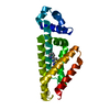
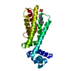
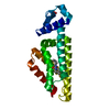
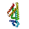
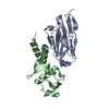
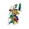
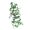
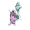
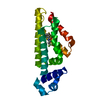
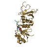
| Title | Human Calcium and Integrin Binding Protein 3 Bound to TMC1 Residues 303-347 | ||||||
 Components Components |
| ||||||
 Keywords Keywords | METAL BINDING/TRANSPORT PROTEIN / EF-hand / Mechanotransduction / hearing / METAL BINDING PROTEIN / METAL BINDING-TRANSPORT PROTEIN complex | ||||||
| Function / homology |  Function and homology information Function and homology informationvestibular reflex / stereocilium tip / auditory receptor cell development / detection of mechanical stimulus involved in sensory perception of sound / mechanosensitive monoatomic ion channel activity / stereocilium / regulation of calcium ion transmembrane transport / calcium ion homeostasis / voltage-gated calcium channel activity / calcium ion transmembrane transport ...vestibular reflex / stereocilium tip / auditory receptor cell development / detection of mechanical stimulus involved in sensory perception of sound / mechanosensitive monoatomic ion channel activity / stereocilium / regulation of calcium ion transmembrane transport / calcium ion homeostasis / voltage-gated calcium channel activity / calcium ion transmembrane transport / calcium channel activity / external side of plasma membrane / calcium ion binding / magnesium ion binding / plasma membrane Similarity search - Function | ||||||
| Biological species |  Homo sapiens (human) Homo sapiens (human) | ||||||
| Method |  X-RAY DIFFRACTION / SYNCHROTRON / MOLECULAR REPLACEMENT / Resolution: 1.84 Å X-RAY DIFFRACTION / SYNCHROTRON / MOLECULAR REPLACEMENT / Resolution: 1.84 Å | ||||||
 Authors Authors | Shapiro, L. / Dionne, G. | ||||||
| Funding support |  United States, 1items United States, 1items
| ||||||
 Citation Citation | Journal: Neuron / Year: 2021 Title: CIB2 and CIB3 are auxiliary subunits of the mechanotransduction channel of hair cells. Authors: Liang, X. / Qiu, X. / Dionne, G. / Cunningham, C.L. / Pucak, M.L. / Peng, G. / Kim, Y.H. / Lauer, A. / Shapiro, L. / Muller, U. | ||||||
| History |
|
- Structure visualization
| Structure viewer | Molecule: MolmilJmol/JSmol |
|---|
- Downloads & links
-Download
| PDBx/mmCIF format | 6wud.cif.gz | 158.2 KB | Display | PDBx/mmCIF format |
|---|---|---|---|---|
| PDB format | pdb6wud.ent.gz | 120 KB | Display | PDB format |
| PDBx/mmJSON format | 6wud.json.gz | Tree view | PDBx/mmJSON format | |
| Others |  Other downloads Other downloads |
-Validation report
| Arichive directory | https://data.pdbj.org/pub/pdb/validation_reports/wu/6wudftp://data.pdbj.org/pub/pdb/validation_reports/wu/6wud | HTTPS FTP |
|---|
-Related structure data
| Related structure data |  6wu5C  6wu7SC C: citing same article ( S: Starting model for refinement |
|---|---|
| Similar structure data |
-Links
 PDBj
PDBj- Assembly
| Deposited unit | 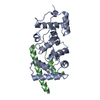
| ||||||||||||
|---|---|---|---|---|---|---|---|---|---|---|---|---|---|
| 1 |
| ||||||||||||
| Unit cell |
|
-Components
| #1: Protein | Mass: 22309.160 Da / Num. of mol.: 1 Source method: isolated from a genetically manipulated source Source: (gene. exp.) Homo sapiens (human) / Gene: CIB3, KIP3 / Production host:  | ||||
|---|---|---|---|---|---|
| #2: Protein | Mass: 6281.816 Da / Num. of mol.: 1 Source method: isolated from a genetically manipulated source Source: (gene. exp.) | ||||
| #3: Chemical |   Mass: 24.305 Da / Num. of mol.: 2 / Source method: obtained synthetically / Formula: Mg Mass: 24.305 Da / Num. of mol.: 2 / Source method: obtained synthetically / Formula: Mg#4: Water | ChemComp-HOH / |  Mass: 18.015 Da / Num. of mol.: 164 / Source method: isolated from a natural source / Formula: H2O Mass: 18.015 Da / Num. of mol.: 164 / Source method: isolated from a natural source / Formula: H2OHas ligand of interest | N | |
-Experimental details
-Experiment
| Experiment | Method: X-RAY DIFFRACTION / Number of used crystals: 1 |
|---|
- Sample preparation
| Crystal | Density Matthews: 2.54 Å3/Da / Density % sol: 51.63 % |
|---|---|
| Crystal grow | Temperature: 293 K / Method: vapor diffusion, hanging drop / pH: 8 / Details: 25% PEG 3350 0.2M Magnesium Chloride |
-Data collection
| Diffraction | Mean temperature: 100 K / Serial crystal experiment: N |
|---|---|
| Diffraction source | Source: SYNCHROTRON / Site: APS / Beamline: 24-ID-C / Wavelength: 1.77 Å |
| Detector | Type: DECTRIS PILATUS 6M-F / Detector: PIXEL / Date: Oct 13, 2019 |
| Radiation | Protocol: SINGLE WAVELENGTH / Monochromatic (M) / Laue (L): M / Scattering type: x-ray |
| Radiation wavelength | Wavelength: 1.77 Å / Relative weight: 1 |
| Reflection | Resolution: 1.838→87.17 Å / Num. obs: 23155 / % possible obs: 90.83 % / Redundancy: 17.8 % / Biso Wilson estimate: 27.65 Å2 / CC1/2: 0.999 / Net I/σ(I): 20.93 |
| Reflection shell | Resolution: 1.838→1.904 Å / Num. unique obs: 1806 / CC1/2: 0.864 / % possible all: 72.18 |
- Processing
| Software |
| ||||||||||||||||||||||||||||||||||||||||||||||||||||||||||||||||||||||||||||||||||||||||||||||||||||||||||||||||||||||||||||||||||||||||||||||||||||||||||||||||||||||||||||||||||||||||||||||||||||
|---|---|---|---|---|---|---|---|---|---|---|---|---|---|---|---|---|---|---|---|---|---|---|---|---|---|---|---|---|---|---|---|---|---|---|---|---|---|---|---|---|---|---|---|---|---|---|---|---|---|---|---|---|---|---|---|---|---|---|---|---|---|---|---|---|---|---|---|---|---|---|---|---|---|---|---|---|---|---|---|---|---|---|---|---|---|---|---|---|---|---|---|---|---|---|---|---|---|---|---|---|---|---|---|---|---|---|---|---|---|---|---|---|---|---|---|---|---|---|---|---|---|---|---|---|---|---|---|---|---|---|---|---|---|---|---|---|---|---|---|---|---|---|---|---|---|---|---|---|---|---|---|---|---|---|---|---|---|---|---|---|---|---|---|---|---|---|---|---|---|---|---|---|---|---|---|---|---|---|---|---|---|---|---|---|---|---|---|---|---|---|---|---|---|---|---|---|---|
| Refinement | Method to determine structure: MOLECULAR REPLACEMENT Starting model: 6WU7 Resolution: 1.84→87.17 Å / SU ML: 0.1683 / Cross valid method: FREE R-VALUE / σ(F): 1.36 / Phase error: 19.7705 Stereochemistry target values: GeoStd + Monomer Library + CDL v1.2
| ||||||||||||||||||||||||||||||||||||||||||||||||||||||||||||||||||||||||||||||||||||||||||||||||||||||||||||||||||||||||||||||||||||||||||||||||||||||||||||||||||||||||||||||||||||||||||||||||||||
| Solvent computation | Shrinkage radii: 0.9 Å / VDW probe radii: 1.11 Å / Solvent model: FLAT BULK SOLVENT MODEL | ||||||||||||||||||||||||||||||||||||||||||||||||||||||||||||||||||||||||||||||||||||||||||||||||||||||||||||||||||||||||||||||||||||||||||||||||||||||||||||||||||||||||||||||||||||||||||||||||||||
| Displacement parameters | Biso mean: 34.95 Å2 | ||||||||||||||||||||||||||||||||||||||||||||||||||||||||||||||||||||||||||||||||||||||||||||||||||||||||||||||||||||||||||||||||||||||||||||||||||||||||||||||||||||||||||||||||||||||||||||||||||||
| Refinement step | Cycle: LAST / Resolution: 1.84→87.17 Å
| ||||||||||||||||||||||||||||||||||||||||||||||||||||||||||||||||||||||||||||||||||||||||||||||||||||||||||||||||||||||||||||||||||||||||||||||||||||||||||||||||||||||||||||||||||||||||||||||||||||
| Refine LS restraints |
| ||||||||||||||||||||||||||||||||||||||||||||||||||||||||||||||||||||||||||||||||||||||||||||||||||||||||||||||||||||||||||||||||||||||||||||||||||||||||||||||||||||||||||||||||||||||||||||||||||||
| LS refinement shell |
|
