 Movie
Movie Controller

[English] 日本語
 Yorodumi
- PDB-6wta: Structure of F420-H2 Dependent Oxidoreductase (FDOR-A) MSMEG_2027... -
+ Open data

- Basic information
| Entry | Database: PDB / ID: 6wta | ||||||
|---|---|---|---|---|---|---|---|
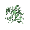
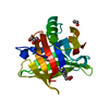
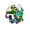
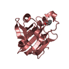
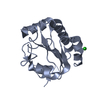
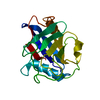
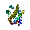
| Title | Structure of F420-H2 Dependent Oxidoreductase (FDOR-A) MSMEG_2027 in complex with F420 | ||||||
 Components Components | Uncharacterized protein | ||||||
 Keywords Keywords | OXIDOREDUCTASE / F420 / Complex / Cofactor | ||||||
| Function / homology | F420H(2)-dependent quinone reductase / F420H(2)-dependent quinone reductase / coenzyme F420 binding / FMN-binding split barrel / oxidoreductase activity / plasma membrane / COENZYME F420-4 / Nitroreductase Function and homology information Function and homology information | ||||||
| Biological species |  Mycolicibacterium smegmatis (bacteria) Mycolicibacterium smegmatis (bacteria) | ||||||
| Method |  X-RAY DIFFRACTION / SYNCHROTRON / MOLECULAR REPLACEMENT / Resolution: 1.67 Å X-RAY DIFFRACTION / SYNCHROTRON / MOLECULAR REPLACEMENT / Resolution: 1.67 Å | ||||||
 Authors Authors | Jackson, C.J. / Antoney, J.P. | ||||||
 Citation Citation | Journal: J.Mol.Biol. / Year: 2025 Title: A F 420 -dependent single domain chemogenetic tool for protein de-dimerization. Authors: Antoney, J. / Kainrath, S. / Dubowsky, J.G. / Ahmed, F.H. / Kang, S.W. / Mackie, E.R.R. / Granado, G.B. / Soares da Costa, T.P. / Jackson, C.J. / Janovjak, H. #1: Journal: Biorxiv / Year: 2022Title: A F420-dependent single domain chemogenetic tool for protein de-dimerization Authors: Antoney, J. / Kainrath, S. / Ahmed, F.H. / Kang, S.W. / Mackie, E.R. / Soares da Costa, T.P. / Jackson, C.J. / Janovjak, H. | ||||||
| History |
|
- Structure visualization
| Structure viewer | Molecule: MolmilJmol/JSmol |
|---|
- Downloads & links
-Download
| PDBx/mmCIF format | 6wta.cif.gz | 106.8 KB | Display | PDBx/mmCIF format |
|---|---|---|---|---|
| PDB format | pdb6wta.ent.gz | 82.5 KB | Display | PDB format |
| PDBx/mmJSON format | 6wta.json.gz | Tree view | PDBx/mmJSON format | |
| Others |  Other downloads Other downloads |
-Validation report
| Summary document | 6wta_validation.pdf.gz | 759.6 KB | Display | wwPDB validaton report |
|---|---|---|---|---|
| Full document | 6wta_full_validation.pdf.gz | 760.8 KB | Display | |
| Data in XML | 6wta_validation.xml.gz | 9.3 KB | Display | |
| Data in CIF | 6wta_validation.cif.gz | 11.9 KB | Display | |
| Arichive directory | https://data.pdbj.org/pub/pdb/validation_reports/wt/6wtaftp://data.pdbj.org/pub/pdb/validation_reports/wt/6wta | HTTPS FTP |
-Related structure data
| Related structure data | 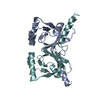 6xriC  4y9iS S: Starting model for refinement C: citing same article ( |
|---|---|
| Similar structure data |


-Links
 PDBj
PDBj- Assembly
| Deposited unit | 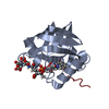
| ||||||||||
|---|---|---|---|---|---|---|---|---|---|---|---|
| 1 | 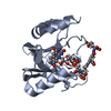
| ||||||||||
| 2 | 
| ||||||||||
| Unit cell |
|
-Components
| #1: Protein | Mass: 16028.034 Da / Num. of mol.: 2 Source method: isolated from a genetically manipulated source Source: (gene. exp.) Mycolicibacterium smegmatis (strain ATCC 700084 / mc(2)155) (bacteria)Strain: ATCC 700084 / mc(2)155 / Gene: MSMEG_2027 / Plasmid: pETMCSIII / Production host: #2: Chemical | ChemComp-UBM / |   Mass: 1031.821 Da / Num. of mol.: 1 / Source method: isolated from a natural source / Formula: C39H50N7O24P / Feature type: SUBJECT OF INVESTIGATION Mass: 1031.821 Da / Num. of mol.: 1 / Source method: isolated from a natural source / Formula: C39H50N7O24P / Feature type: SUBJECT OF INVESTIGATION#3: Water | ChemComp-HOH / |  Mass: 18.015 Da / Num. of mol.: 87 / Source method: isolated from a natural source / Formula: H2O Mass: 18.015 Da / Num. of mol.: 87 / Source method: isolated from a natural source / Formula: H2OHas ligand of interest | Y | Has protein modification | N | |
|---|
-Experimental details
-Experiment
| Experiment | Method: X-RAY DIFFRACTION / Number of used crystals: 1 |
|---|
- Sample preparation
| Crystal | Density Matthews: 2.3 Å3/Da / Density % sol: 46.5 % |
|---|---|
| Crystal grow | Temperature: 291.15 K / Method: vapor diffusion, hanging drop / pH: 3.4 Details: 0.07 M citrate, 0.03 M bis-tris propane, 16% PEG 3350 |
-Data collection
| Diffraction | Mean temperature: 100 K / Serial crystal experiment: N |
|---|---|
| Diffraction source | Source: SYNCHROTRON / Site: Australian Synchrotron  / Beamline: MX2 / Wavelength: 0.953729987144 Å / Beamline: MX2 / Wavelength: 0.953729987144 Å |
| Detector | Type: DECTRIS EIGER X 16M / Detector: PIXEL / Date: Nov 2, 2017 |
| Radiation | Protocol: SINGLE WAVELENGTH / Monochromatic (M) / Laue (L): M / Scattering type: x-ray |
| Radiation wavelength | Wavelength: 0.953729987144 Å / Relative weight: 1 |
| Reflection | Resolution: 1.67→32.68 Å / Num. obs: 17596 / % possible obs: 99.8 % / Redundancy: 10.6 % / Biso Wilson estimate: 19.79 Å2 / CC1/2: 0.996 / Rmerge(I) obs: 0.173 / Rpim(I) all: 0.078 / Rrim(I) all: 0.183 / Χ2: 0.93 / Net I/σ(I): 11.8 |
| Reflection shell | Resolution: 1.67→1.7 Å / Redundancy: 10.1 % / Rmerge(I) obs: 1.556 / Mean I/σ(I) obs: 1.9 / Num. unique obs: 886 / CC1/2: 0.91 / Rpim(I) all: 0.511 / Χ2: 1.02 / % possible all: 99.9 |
- Processing
| Software |
| |||||||||||||||||||||||||||||||||||||||||||||||||||||||||||||||||||||||||||||||||||||||||||||||||||||||||||||||||||||||||||||
|---|---|---|---|---|---|---|---|---|---|---|---|---|---|---|---|---|---|---|---|---|---|---|---|---|---|---|---|---|---|---|---|---|---|---|---|---|---|---|---|---|---|---|---|---|---|---|---|---|---|---|---|---|---|---|---|---|---|---|---|---|---|---|---|---|---|---|---|---|---|---|---|---|---|---|---|---|---|---|---|---|---|---|---|---|---|---|---|---|---|---|---|---|---|---|---|---|---|---|---|---|---|---|---|---|---|---|---|---|---|---|---|---|---|---|---|---|---|---|---|---|---|---|---|---|---|---|
| Refinement | Method to determine structure: MOLECULAR REPLACEMENT Starting model: 4Y9I Resolution: 1.67→32.68 Å / SU ML: 0.21 / Cross valid method: FREE R-VALUE / σ(F): 1.34 / Phase error: 36.33 / Stereochemistry target values: ML
| |||||||||||||||||||||||||||||||||||||||||||||||||||||||||||||||||||||||||||||||||||||||||||||||||||||||||||||||||||||||||||||
| Solvent computation | Shrinkage radii: 0.9 Å / VDW probe radii: 1.11 Å / Solvent model: FLAT BULK SOLVENT MODEL | |||||||||||||||||||||||||||||||||||||||||||||||||||||||||||||||||||||||||||||||||||||||||||||||||||||||||||||||||||||||||||||
| Refinement step | Cycle: LAST / Resolution: 1.67→32.68 Å
| |||||||||||||||||||||||||||||||||||||||||||||||||||||||||||||||||||||||||||||||||||||||||||||||||||||||||||||||||||||||||||||
| Refine LS restraints |
| |||||||||||||||||||||||||||||||||||||||||||||||||||||||||||||||||||||||||||||||||||||||||||||||||||||||||||||||||||||||||||||
| LS refinement shell |
| |||||||||||||||||||||||||||||||||||||||||||||||||||||||||||||||||||||||||||||||||||||||||||||||||||||||||||||||||||||||||||||
| Refinement TLS params. | Method: refined / Refine-ID: X-RAY DIFFRACTION
| |||||||||||||||||||||||||||||||||||||||||||||||||||||||||||||||||||||||||||||||||||||||||||||||||||||||||||||||||||||||||||||
| Refinement TLS group |
|
