 Movie
Movie Controller

[English] 日本語
 Yorodumi
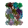
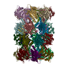
- PDB-6wnk: Macrocyclic peptides TDI5575 that selectively inhibit the Mycobac... -
+ Open data

- Basic information
| Entry | Database: PDB / ID: 6wnk | ||||||
|---|---|---|---|---|---|---|---|
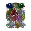
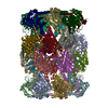
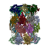
| Title | Macrocyclic peptides TDI5575 that selectively inhibit the Mycobacterium tuberculosis proteasome | ||||||
 Components Components | (Proteasome subunit ...) x 2 | ||||||
 Keywords Keywords | HYDROLASE/HYDROLASE INHIBITOR / Mycobacterium tuberculosis / proteasome inhibitor / Macrocyclic peptides / HYDROLASE / HYDROLASE-HYDROLASE INHIBITOR complex | ||||||
| Function / homology |  Function and homology information Function and homology informationproteasome endopeptidase complex / proteasome core complex, beta-subunit complex / threonine-type endopeptidase activity / proteasome core complex, alpha-subunit complex / proteasomal protein catabolic process / modification-dependent protein catabolic process / cytoplasm Similarity search - Function | ||||||
| Biological species |   Mycobacterium tuberculosis (bacteria) Mycobacterium tuberculosis (bacteria) | ||||||
| Method |  X-RAY DIFFRACTION / SYNCHROTRON / MOLECULAR REPLACEMENT / Resolution: 2.28 Å X-RAY DIFFRACTION / SYNCHROTRON / MOLECULAR REPLACEMENT / Resolution: 2.28 Å | ||||||
 Authors Authors | Hsu, H.C. / Li, H. | ||||||
| Funding support |  United States, 1items United States, 1items
| ||||||
 Citation Citation | Journal: J.Med.Chem. / Year: 2021 Title: Macrocyclic Peptides that Selectively Inhibit the Mycobacterium tuberculosis Proteasome. Authors: Zhang, H. / Hsu, H.C. / Kahne, S.C. / Hara, R. / Zhan, W. / Jiang, X. / Burns-Huang, K. / Ouellette, T. / Imaeda, T. / Okamoto, R. / Kawasaki, M. / Michino, M. / Wong, T.T. / Toita, A. / ...Authors: Zhang, H. / Hsu, H.C. / Kahne, S.C. / Hara, R. / Zhan, W. / Jiang, X. / Burns-Huang, K. / Ouellette, T. / Imaeda, T. / Okamoto, R. / Kawasaki, M. / Michino, M. / Wong, T.T. / Toita, A. / Yukawa, T. / Moraca, F. / Vendome, J. / Saha, P. / Sato, K. / Aso, K. / Ginn, J. / Meinke, P.T. / Foley, M. / Nathan, C.F. / Darwin, K.H. / Li, H. / Lin, G. | ||||||
| History |
|
- Structure visualization
| Structure viewer | Molecule: MolmilJmol/JSmol |
|---|
- Downloads & links
-Download
| PDBx/mmCIF format | 6wnk.cif.gz | 1.2 MB | Display | PDBx/mmCIF format |
|---|---|---|---|---|
| PDB format | pdb6wnk.ent.gz | 969.5 KB | Display | PDB format |
| PDBx/mmJSON format | 6wnk.json.gz | Tree view | PDBx/mmJSON format | |
| Others |  Other downloads Other downloads |
-Validation report
| Arichive directory | https://data.pdbj.org/pub/pdb/validation_reports/wn/6wnkftp://data.pdbj.org/pub/pdb/validation_reports/wn/6wnk | HTTPS FTP |
|---|
-Related structure data
| Related structure data | 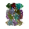 5trgS S: Starting model for refinement |
|---|---|
| Similar structure data |


-Links
 PDBj
PDBj


- Assembly
| Deposited unit | 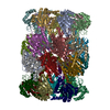
| ||||||||||||
|---|---|---|---|---|---|---|---|---|---|---|---|---|---|
| 1 |
| ||||||||||||
| Unit cell |
|
-Components
-Proteasome subunit ... , 2 types, 28 molecules ABCDEFGOPQRSTUHIJKLMNVWXYZab
| #1: Protein | Mass: 25971.975 Da / Num. of mol.: 14 Source method: isolated from a genetically manipulated source Source: (gene. exp.) Mycobacterium tuberculosis (bacteria) / Gene: prcA, MRA_2124 / Plasmid: pACYCDuet / Production host: References: UniProt: A5U4D5, proteasome endopeptidase complex #2: Protein | Mass: 25274.264 Da / Num. of mol.: 14 Source method: isolated from a genetically manipulated source Source: (gene. exp.) Mycobacterium tuberculosis (bacteria) / Gene: prcB, MRA_2125 / Plasmid: PACYCDuet / Production host: References: UniProt: A5U4D6, proteasome endopeptidase complex |
|---|
-Non-polymers , 4 types, 1212 molecules 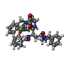


| #3: Chemical | ChemComp-U5Y / (  Type: Cyclic peptide / Class: Inhibitor / Mass: 662.749 Da / Num. of mol.: 14 / Source method: obtained synthetically / Formula: C39H39FN4O5 / Feature type: SUBJECT OF INVESTIGATION / References: Macrocyclic peptide TDI5575 Type: Cyclic peptide / Class: Inhibitor / Mass: 662.749 Da / Num. of mol.: 14 / Source method: obtained synthetically / Formula: C39H39FN4O5 / Feature type: SUBJECT OF INVESTIGATION / References: Macrocyclic peptide TDI5575#4: Chemical | ChemComp-CIT /  Mass: 192.124 Da / Num. of mol.: 14 / Source method: obtained synthetically / Formula: C6H8O7 Mass: 192.124 Da / Num. of mol.: 14 / Source method: obtained synthetically / Formula: C6H8O7#5: Chemical | ChemComp-DMF /  Mass: 73.094 Da / Num. of mol.: 8 / Source method: obtained synthetically / Formula: C3H7NO Mass: 73.094 Da / Num. of mol.: 8 / Source method: obtained synthetically / Formula: C3H7NO#6: Water | ChemComp-HOH / | Mass: 18.015 Da / Num. of mol.: 1176 / Source method: isolated from a natural source / Formula: H2O |
|---|
-Details
| Has ligand of interest | Y |
|---|
-Experimental details
-Experiment
| Experiment | Method: X-RAY DIFFRACTION / Number of used crystals: 1 |
|---|
- Sample preparation
| Crystal | Density Matthews: 2.62 Å3/Da / Density % sol: 52.99 % |
|---|---|
| Crystal grow | Temperature: 277 K / Method: vapor diffusion, hanging drop / pH: 6.2 / Details: 60mM sodium citrate (pH 6.2), 15% PEG-3350 |
-Data collection
| Diffraction | Mean temperature: 100 K / Serial crystal experiment: N |
|---|---|
| Diffraction source | Source: SYNCHROTRON / Site: APS / Beamline: 21-ID-D / Wavelength: 1.07812 Å |
| Detector | Type: DECTRIS EIGER X 9M / Detector: PIXEL / Date: Apr 14, 2017 |
| Radiation | Protocol: SINGLE WAVELENGTH / Monochromatic (M) / Laue (L): M / Scattering type: x-ray |
| Radiation wavelength | Wavelength: 1.07812 Å / Relative weight: 1 |
| Reflection | Resolution: 2.28→92.97 Å / Num. obs: 332027 / % possible obs: 98.2 % / Redundancy: 7 % / Biso Wilson estimate: 42.06 Å2 / CC1/2: 0.997 / Rmerge(I) obs: 0.11 / Net I/σ(I): 10.7 |
| Reflection shell | Resolution: 2.28→2.4 Å / Redundancy: 7.2 % / Rmerge(I) obs: 0.85 / Mean I/σ(I) obs: 2.3 / Num. unique obs: 48823 / CC1/2: 0.724 / % possible all: 99.2 |
- Processing
| Software |
| ||||||||||||||||||||||||
|---|---|---|---|---|---|---|---|---|---|---|---|---|---|---|---|---|---|---|---|---|---|---|---|---|---|
| Refinement | Method to determine structure: MOLECULAR REPLACEMENT Starting model: 5TRG Resolution: 2.28→87.59 Å / Cross valid method: FREE R-VALUE
| ||||||||||||||||||||||||
| Displacement parameters | Biso mean: 53.04 Å2 | ||||||||||||||||||||||||
| Refinement step | Cycle: LAST / Resolution: 2.28→87.59 Å
| ||||||||||||||||||||||||
| Refine LS restraints |
|
