+ データを開く
- 基本情報
基本情報
| 登録情報 | データベース: PDB / ID: 6wkk | ||||||
|---|---|---|---|---|---|---|---|
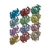
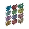
| タイトル | Phage G gp27 major capsid proteins and gp26 decoration proteins | ||||||
 要素 要素 |
| ||||||
 キーワード キーワード | VIRUS / phage G / major capsid protein / decoration protein / capsid / icosahedral / gp26 / gp27 | ||||||
| 機能・相同性 | : / Phage capsid / Phage capsid family / Gp26 / Gp27 機能・相同性情報 機能・相同性情報 | ||||||
| 生物種 |  Bacillus virus G (ウイルス) Bacillus virus G (ウイルス) | ||||||
| 手法 | 電子顕微鏡法 / 単粒子再構成法 / クライオ電子顕微鏡法 / 解像度: 6.1 Å | ||||||
 データ登録者 データ登録者 | Monroe, L. / Gonzalez, B. / Jiang, W. / Kihara, D. | ||||||
 引用 引用 |  ジャーナル: J Mol Biol / 年: 2020 ジャーナル: J Mol Biol / 年: 2020タイトル: Phage G Structure at 6.1 Å Resolution, Condensed DNA, and Host Identity Revision to a Lysinibacillus. 著者: Brenda González / Lyman Monroe / Kunpeng Li / Rui Yan / Elena Wright / Thomas Walter / Daisuke Kihara / Susan T Weintraub / Julie A Thomas / Philip Serwer / Wen Jiang /  要旨: Phage G has the largest capsid and genome of any known propagated phage. Many aspects of its structure, assembly, and replication have not been elucidated. Herein, we present the dsDNA-packed and ...Phage G has the largest capsid and genome of any known propagated phage. Many aspects of its structure, assembly, and replication have not been elucidated. Herein, we present the dsDNA-packed and empty phage G capsid at 6.1 and 9 Å resolution, respectively, using cryo-EM for structure determination and mass spectrometry for protein identification. The major capsid protein, gp27, is identified and found to share the HK97-fold universally conserved in all previously solved dsDNA phages. Trimers of the decoration protein, gp26, sit on the 3-fold axes and are thought to enhance the interactions of the hexameric capsomeres of gp27, for other phages encoding decoration proteins. Phage G's decoration protein is longer than what has been reported in other phages, and we suspect the extra interaction surface area helps stabilize the capsid. We identified several additional capsid proteins, including a candidate for the prohead protease responsible for processing gp27. Furthermore, cryo-EM reveals a range of partially full, condensed DNA densities that appear to have no contact with capsid shell. Three analyses confirm that the phage G host is a Lysinibacillus, and not Bacillus megaterium: identity of host proteins in our mass spectrometry analyses, genome sequence of the phage G host, and host range of phage G. | ||||||
| 履歴 |
|
- 構造の表示
構造の表示
| ムービー |
 ムービービューア ムービービューア |
|---|---|
| 構造ビューア | 分子:  Molmil Molmil Jmol/JSmol Jmol/JSmol |
- ダウンロードとリンク
- ダウンロード
| PDBx/mmCIF形式 |  6wkk.cif.gz 6wkk.cif.gz | 704.3 KB | 表示 |  PDBx/mmCIF形式 PDBx/mmCIF形式 |
|---|---|---|---|---|
| PDB形式 |  pdb6wkk.ent.gz pdb6wkk.ent.gz | 560.5 KB | 表示 |  PDB形式 PDB形式 |
| PDBx/mmJSON形式 |  6wkk.json.gz 6wkk.json.gz | ツリー表示 |  PDBx/mmJSON形式 PDBx/mmJSON形式 | |
| その他 |  その他のダウンロード その他のダウンロード |
-検証レポート
| 文書・要旨 |  6wkk_validation.pdf.gz 6wkk_validation.pdf.gz | 524.4 KB | 表示 |  wwPDB検証レポート wwPDB検証レポート |
|---|---|---|---|---|
| 文書・詳細版 |  6wkk_full_validation.pdf.gz 6wkk_full_validation.pdf.gz | 703.9 KB | 表示 | |
| XML形式データ |  6wkk_validation.xml.gz 6wkk_validation.xml.gz | 95 KB | 表示 | |
| CIF形式データ |  6wkk_validation.cif.gz 6wkk_validation.cif.gz | 143.9 KB | 表示 | |
| アーカイブディレクトリ |  https://data.pdbj.org/pub/pdb/validation_reports/wk/6wkk https://data.pdbj.org/pub/pdb/validation_reports/wk/6wkk ftp://data.pdbj.org/pub/pdb/validation_reports/wk/6wkk ftp://data.pdbj.org/pub/pdb/validation_reports/wk/6wkk | HTTPS FTP |
-関連構造データ
- リンク
リンク
- 集合体
集合体
| 登録構造単位 | 
|
|---|---|
| 1 |
|
- 要素
要素
| #1: タンパク質 | 分子量: 30981.451 Da / 分子数: 6 / 由来タイプ: 天然 / 由来: (天然)  Bacillus virus G (ウイルス) / 参照: UniProt: G3MB97 Bacillus virus G (ウイルス) / 参照: UniProt: G3MB97#2: タンパク質 | 分子量: 15763.595 Da / 分子数: 18 / 由来タイプ: 天然 / 由来: (天然)  Bacillus virus G (ウイルス) / 参照: UniProt: G3MB96 Bacillus virus G (ウイルス) / 参照: UniProt: G3MB96 |
|---|
-実験情報
-実験
| 実験 | 手法: 電子顕微鏡法 |
|---|---|
| EM実験 | 試料の集合状態: PARTICLE / 3次元再構成法: 単粒子再構成法 |
- 試料調製
試料調製
| 構成要素 | 名称: Bacillus virus G / タイプ: VIRUS / Entity ID: all / 由来: NATURAL |
|---|---|
| 由来(天然) | 生物種:  Bacillus virus G (ウイルス) Bacillus virus G (ウイルス) |
| ウイルスについての詳細 | 中空か: NO / エンベロープを持つか: NO / 単離: SPECIES / タイプ: VIRION |
| ウイルス殻 | 直径: 1600 nm / 三角数 (T数): 52 |
| 緩衝液 | pH: 7.4 |
| 試料 | 包埋: NO / シャドウイング: NO / 染色: NO / 凍結: YES 詳細: In brief, phage G was grown in agarose overlays, concentrated by centrifugal pelleting and subjected to rate zonal centrifugation in a sucrose gradient. After recovery from the gradient, ...詳細: In brief, phage G was grown in agarose overlays, concentrated by centrifugal pelleting and subjected to rate zonal centrifugation in a sucrose gradient. After recovery from the gradient, phage G was stored in 0.01 M Tris-Cl (pH 7.4), 0.01 M MgSO4, 6% polyethylene glycol MW 3350. |
| 急速凍結 | 装置: GATAN CRYOPLUNGE 3 / 凍結剤: ETHANE / 湿度: 65 % / 凍結前の試料温度: 298 K |
- 電子顕微鏡撮影
| 実験機器 |  モデル: Titan Krios / 画像提供: FEI Company |
|---|---|
| 顕微鏡 | モデル: FEI TITAN KRIOS |
| 電子銃 | 電子線源:  FIELD EMISSION GUN / 加速電圧: 300 kV / 照射モード: FLOOD BEAM FIELD EMISSION GUN / 加速電圧: 300 kV / 照射モード: FLOOD BEAM |
| 電子レンズ | モード: BRIGHT FIELD |
| 撮影 | 電子線照射量: 14.5 e/Å2 フィルム・検出器のモデル: GATAN K2 SUMMIT (4k x 4k) 実像数: 375 |
- 解析
解析
| EMソフトウェア |
| ||||||||||||||||||||||||||||||||
|---|---|---|---|---|---|---|---|---|---|---|---|---|---|---|---|---|---|---|---|---|---|---|---|---|---|---|---|---|---|---|---|---|---|
| CTF補正 | タイプ: PHASE FLIPPING AND AMPLITUDE CORRECTION | ||||||||||||||||||||||||||||||||
| 粒子像の選択 | 選択した粒子像数: 2564 | ||||||||||||||||||||||||||||||||
| 3次元再構成 | 解像度: 6.1 Å / 解像度の算出法: FSC 0.143 CUT-OFF / 粒子像の数: 2564 詳細: Phage G's capsid was reconstructed with icosahedral symmetry 対称性のタイプ: POINT |
 ムービー
ムービー コントローラー


 PDBj
PDBj

