 Movie
Movie Controller

[English] 日本語
 Yorodumi
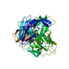
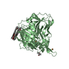
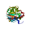
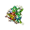
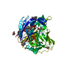
- PDB-6wcl: Crystal structure of Laccase from Thermus thermophilus HB27 with ... -
+ Open data

- Basic information
| Entry | Database: PDB / ID: 6wcl | ||||||
|---|---|---|---|---|---|---|---|
| Title | Crystal structure of Laccase from Thermus thermophilus HB27 with an open conformation of beta-hairpin (Average deposited dose 12.91 MGy) | ||||||
 Components Components | Laccase | ||||||
 Keywords Keywords | OXIDOREDUCTASE / open conformation / multicopper oxidase / laccase | ||||||
| Function / homology |  Function and homology information Function and homology informationhydroquinone:oxygen oxidoreductase activity / laccase / copper ion binding Similarity search - Function | ||||||
| Biological species |   Thermus thermophilus (bacteria) Thermus thermophilus (bacteria) | ||||||
| Method |  X-RAY DIFFRACTION / SYNCHROTRON / MOLECULAR REPLACEMENT / molecular replacement / Resolution: 1.7 Å X-RAY DIFFRACTION / SYNCHROTRON / MOLECULAR REPLACEMENT / molecular replacement / Resolution: 1.7 Å | ||||||
 Authors Authors | Miranda-Blancas, R. / Rudino-Pinera, E. | ||||||
 Citation Citation | Journal: To Be Published Title: Dynamic behavior of beta-hairpin loop at laccase of Thermus thermophilus at 1.7 angstroms resolution Authors: Miranda-Blancas, R. / Rudino-Pinera, E. | ||||||
| History |
|
- Structure visualization
| Structure viewer | Molecule: MolmilJmol/JSmol |
|---|
- Downloads & links
-Download
| PDBx/mmCIF format | 6wcl.cif.gz | 113.2 KB | Display | PDBx/mmCIF format |
|---|---|---|---|---|
| PDB format | pdb6wcl.ent.gz | 83.1 KB | Display | PDB format |
| PDBx/mmJSON format | 6wcl.json.gz | Tree view | PDBx/mmJSON format | |
| Others |  Other downloads Other downloads |
-Validation report
| Arichive directory | https://data.pdbj.org/pub/pdb/validation_reports/wc/6wclftp://data.pdbj.org/pub/pdb/validation_reports/wc/6wcl | HTTPS FTP |
|---|
-Related structure data
| Related structure data |  6wcgC  6wcmC 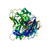 6wcnC 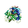 6wcpC  2xu9S S: Starting model for refinement C: citing same article ( |
|---|---|
| Similar structure data |


-Links
 PDBj
PDBj- Assembly
| Deposited unit | 
| ||||||||
|---|---|---|---|---|---|---|---|---|---|
| 1 |
| ||||||||
| Unit cell |
|
-Components
| #1: Protein | Mass: 48848.512 Da / Num. of mol.: 1 Source method: isolated from a genetically manipulated source Source: (gene. exp.) Thermus thermophilus (strain HB27 / ATCC BAA-163 / DSM 7039) (bacteria)Strain: HB27 / ATCC BAA-163 / DSM 7039 / Gene: TT_C1370 / Production host: | ||||||
|---|---|---|---|---|---|---|---|
| #2: Chemical |   Mass: 63.546 Da / Num. of mol.: 2 / Source method: obtained synthetically / Formula: Cu Mass: 63.546 Da / Num. of mol.: 2 / Source method: obtained synthetically / Formula: Cu#3: Chemical | ChemComp-1PE / |   Mass: 238.278 Da / Num. of mol.: 1 / Source method: obtained synthetically / Formula: C10H22O6 / Comment: precipitant*YM Mass: 238.278 Da / Num. of mol.: 1 / Source method: obtained synthetically / Formula: C10H22O6 / Comment: precipitant*YM#4: Water | ChemComp-HOH / |  Mass: 18.015 Da / Num. of mol.: 371 / Source method: isolated from a natural source / Formula: H2O Mass: 18.015 Da / Num. of mol.: 371 / Source method: isolated from a natural source / Formula: H2OHas ligand of interest | N | |
-Experimental details
-Experiment
| Experiment | Method: X-RAY DIFFRACTION / Number of used crystals: 1 |
|---|
- Sample preparation
| Crystal | Density Matthews: 2.44 Å3/Da / Density % sol: 49.58 % / Description: triangular shaped sheets |
|---|---|
| Crystal grow | Temperature: 277.15 K / Method: microbatch / pH: 5.5 Details: 20% PEG 4000, 20% 2-propanol, 100 mM citrate buffer PH range: 5.5-5.6 |
-Data collection
| Diffraction | Mean temperature: 100 K / Serial crystal experiment: N | ||||||||||||||||||||||||||||||||||||||||||||||||||||||||||||||||||||||||||||||||||||||||
|---|---|---|---|---|---|---|---|---|---|---|---|---|---|---|---|---|---|---|---|---|---|---|---|---|---|---|---|---|---|---|---|---|---|---|---|---|---|---|---|---|---|---|---|---|---|---|---|---|---|---|---|---|---|---|---|---|---|---|---|---|---|---|---|---|---|---|---|---|---|---|---|---|---|---|---|---|---|---|---|---|---|---|---|---|---|---|---|---|---|
| Diffraction source | Source: SYNCHROTRON / Site: APS  / Beamline: 19-ID / Wavelength: 1.1949 Å / Beamline: 19-ID / Wavelength: 1.1949 Å | ||||||||||||||||||||||||||||||||||||||||||||||||||||||||||||||||||||||||||||||||||||||||
| Detector | Type: ADSC QUANTUM 315 / Detector: CCD / Date: Apr 17, 2018 / Details: Sagittal focusing 2nd crystal horizontal focusing | ||||||||||||||||||||||||||||||||||||||||||||||||||||||||||||||||||||||||||||||||||||||||
| Radiation | Monochromator: Si (111) Rosenbaum-Rock double-crystal monochromator: liquid nitrogen cooled Protocol: SINGLE WAVELENGTH / Monochromatic (M) / Laue (L): M / Scattering type: x-ray | ||||||||||||||||||||||||||||||||||||||||||||||||||||||||||||||||||||||||||||||||||||||||
| Radiation wavelength | Wavelength: 1.1949 Å / Relative weight: 1 | ||||||||||||||||||||||||||||||||||||||||||||||||||||||||||||||||||||||||||||||||||||||||
| Reflection | Resolution: 1.7→39.24 Å / Num. obs: 51854 / % possible obs: 99.97 % / Redundancy: 1.9 % / Biso Wilson estimate: 19.11 Å2 / CC1/2: 0.997 / CC star: 0.999 / Rmerge(I) obs: 0.048 / Rpim(I) all: 0.048 / Rrim(I) all: 0.068 / Net I/σ(I): 7.93 | ||||||||||||||||||||||||||||||||||||||||||||||||||||||||||||||||||||||||||||||||||||||||
| Reflection shell | Diffraction-ID: 1 / % possible all: 100
|
-Phasing
| Phasing | Method: molecular replacement |
|---|
- Processing
| Software |
| ||||||||||||||||||||||||||||||||||||||||||||||||||||||||||||||||||||||||||||||||||||||||||||||||||||||||||||||||||||||||
|---|---|---|---|---|---|---|---|---|---|---|---|---|---|---|---|---|---|---|---|---|---|---|---|---|---|---|---|---|---|---|---|---|---|---|---|---|---|---|---|---|---|---|---|---|---|---|---|---|---|---|---|---|---|---|---|---|---|---|---|---|---|---|---|---|---|---|---|---|---|---|---|---|---|---|---|---|---|---|---|---|---|---|---|---|---|---|---|---|---|---|---|---|---|---|---|---|---|---|---|---|---|---|---|---|---|---|---|---|---|---|---|---|---|---|---|---|---|---|---|---|---|
| Refinement | Method to determine structure: MOLECULAR REPLACEMENT Starting model: 2XU9 Resolution: 1.7→39.24 Å / SU ML: 0.26 / Cross valid method: THROUGHOUT / σ(F): 1.35 / Phase error: 25.38
| ||||||||||||||||||||||||||||||||||||||||||||||||||||||||||||||||||||||||||||||||||||||||||||||||||||||||||||||||||||||||
| Solvent computation | Shrinkage radii: 0.9 Å / VDW probe radii: 1.11 Å | ||||||||||||||||||||||||||||||||||||||||||||||||||||||||||||||||||||||||||||||||||||||||||||||||||||||||||||||||||||||||
| Displacement parameters | Biso max: 120.84 Å2 / Biso mean: 24.2195 Å2 / Biso min: 7.15 Å2 | ||||||||||||||||||||||||||||||||||||||||||||||||||||||||||||||||||||||||||||||||||||||||||||||||||||||||||||||||||||||||
| Refinement step | Cycle: final / Resolution: 1.7→39.24 Å
| ||||||||||||||||||||||||||||||||||||||||||||||||||||||||||||||||||||||||||||||||||||||||||||||||||||||||||||||||||||||||
| LS refinement shell | Refine-ID: X-RAY DIFFRACTION / Rfactor Rfree error: 0 / Total num. of bins used: 19 / % reflection obs: 100 %
|
