 Movie
Movie Controller

[English] 日本語
 Yorodumi
- PDB-4ai7: Crystal structure of Laccase from Thermus thermophilus HB27 compl... -
+ Open data

- Basic information
| Entry | Database: PDB / ID: 4ai7 | ||||||
|---|---|---|---|---|---|---|---|
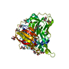
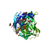
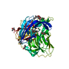
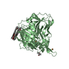
| Title | Crystal structure of Laccase from Thermus thermophilus HB27 complexed with Hg, crystal of the apoenzyme soaked for 2 h in 5 mM HgCl2 at 278 K. | ||||||
 Components Components | LACCASE-LIKE PROTEIN | ||||||
 Keywords Keywords | OXIDOREDUCTASE / MULTICOPPER OXIDASE | ||||||
| Function / homology |  Function and homology information Function and homology informationhydroquinone:oxygen oxidoreductase activity / laccase / oxidoreductase activity / copper ion binding Similarity search - Function | ||||||
| Biological species |   THERMUS THERMOPHILUS (bacteria) THERMUS THERMOPHILUS (bacteria) | ||||||
| Method |  X-RAY DIFFRACTION / SYNCHROTRON / MOLECULAR REPLACEMENT / Resolution: 1.7 Å X-RAY DIFFRACTION / SYNCHROTRON / MOLECULAR REPLACEMENT / Resolution: 1.7 Å | ||||||
 Authors Authors | Serrano-Posada, H. / Rudino-Pinera, E. | ||||||
 Citation Citation | Journal: Acta Crystallogr.,Sect.D / Year: 2015 Title: X-Ray-Induced Catalytic Active-Site Reduction of a Multicopper Oxidase: Structural Insights Into the Proton-Relay Mechanism and O2-Reduction States. Authors: Serrano-Posada, H. / Centeno-Leija, S. / Rojas-Trejo, S.P. / Rodriguez-Almazan, C. / Stojanoff, V. / Rudino-Pinera, E. | ||||||
| History |
|
- Structure visualization
| Structure viewer | Molecule: MolmilJmol/JSmol |
|---|
- Downloads & links
-Download
| PDBx/mmCIF format | 4ai7.cif.gz | 125.5 KB | Display | PDBx/mmCIF format |
|---|---|---|---|---|
| PDB format | pdb4ai7.ent.gz | 97 KB | Display | PDB format |
| PDBx/mmJSON format | 4ai7.json.gz | Tree view | PDBx/mmJSON format | |
| Others |  Other downloads Other downloads |
-Validation report
| Arichive directory | https://data.pdbj.org/pub/pdb/validation_reports/ai/4ai7ftp://data.pdbj.org/pub/pdb/validation_reports/ai/4ai7 | HTTPS FTP |
|---|
-Related structure data
| Related structure data |  2xu9SC  2xuwC  2xvbC  2yaeC  2yafC  2yahC  2yamC  2yaoC  2yapC  2yaqC  2yarC S: Starting model for refinement C: citing same article ( |
|---|---|
| Similar structure data |


-Links
 PDBj
PDBj


- Assembly
| Deposited unit | 
| ||||||||||||
|---|---|---|---|---|---|---|---|---|---|---|---|---|---|
| 1 |
| ||||||||||||
| Unit cell |
| ||||||||||||
| Components on special symmetry positions |
|
-Components
| #1: Protein | Mass: 48791.457 Da / Num. of mol.: 1 Source method: isolated from a genetically manipulated source Details: THE PRESENCE OF AN ISOLEUCINE AT THE POSITION 53 IS STRONGLY SUPPORTED BY THE ELECTRON DENSITY Source: (gene. exp.) THERMUS THERMOPHILUS (bacteria) / Strain: HB27 / Production host: | ||||||
|---|---|---|---|---|---|---|---|
| #2: Chemical | ChemComp-HG /   Mass: 200.590 Da / Num. of mol.: 5 / Source method: obtained synthetically / Formula: Hg Mass: 200.590 Da / Num. of mol.: 5 / Source method: obtained synthetically / Formula: Hg#3: Chemical | ChemComp-MPD / (   Mass: 118.174 Da / Num. of mol.: 16 / Source method: obtained synthetically / Formula: C6H14O2 / Comment: precipitant*YM Mass: 118.174 Da / Num. of mol.: 16 / Source method: obtained synthetically / Formula: C6H14O2 / Comment: precipitant*YM#4: Water | ChemComp-HOH / |  Mass: 18.015 Da / Num. of mol.: 504 / Source method: isolated from a natural source / Formula: H2O Mass: 18.015 Da / Num. of mol.: 504 / Source method: isolated from a natural source / Formula: H2OSequence details | THE SEQUENCE AT THE UNIPROT DEPOSIT Q4H436 POSITION 53 IS OCCUPIED BY A LEUCINE BUT THE ELECTRON ...THE SEQUENCE AT THE UNIPROT DEPOSIT Q4H436 POSITION 53 IS OCCUPIED BY A LEUCINE BUT THE ELECTRON DENSITY CLEARLY SUPPORT THE PRESENCE OF AN ISOLEUCINE | |
-Experimental details
-Experiment
| Experiment | Method: X-RAY DIFFRACTION / Number of used crystals: 1 |
|---|
- Sample preparation
| Crystal | Density Matthews: 2.59 Å3/Da / Density % sol: 52.47 % / Description: NONE |
|---|---|
| Crystal grow | pH: 7.5 Details: 0.1 M HEPES PH 7.5, 70 % MPD BEFORE DATA COLLECTION THE CRYSTAL WAS SOAKED FOR 2 HOURS IN 5 MM HGCL2 |
-Data collection
| Diffraction | Mean temperature: 100 K |
|---|---|
| Diffraction source | Source: SYNCHROTRON / Site: NSLS  / Beamline: X6A / Wavelength: 0.9795 / Beamline: X6A / Wavelength: 0.9795 |
| Detector | Type: ADSC QUANTUM 210 / Detector: CCD / Date: Apr 2, 2011 Details: DOUBLE CRYSTAL CHANNEL CUT, SI(111), 1M LONG RH COATED TOROIDAL MIRROR FOR VERTICAL AND HORIZONTAL FOCUSING. |
| Radiation | Protocol: SINGLE WAVELENGTH / Monochromatic (M) / Laue (L): M / Scattering type: x-ray |
| Radiation wavelength | Wavelength: 0.9795 Å / Relative weight: 1 |
| Reflection | Resolution: 1.7→28 Å / Num. obs: 53278 / % possible obs: 97.9 % / Observed criterion σ(I): 0 / Redundancy: 4.6 % / Biso Wilson estimate: 14.14 Å2 / Rmerge(I) obs: 0.06 / Net I/σ(I): 18.4 |
| Reflection shell | Resolution: 1.7→1.8 Å / Redundancy: 4.7 % / Rmerge(I) obs: 0.28 / Mean I/σ(I) obs: 5.1 / % possible all: 97.5 |
- Processing
| Software |
| ||||||||||||||||||||||||||||||||||||||||||||||||||||||||||||||||||||||||||||||||||||||||||||||||||||||||||||||||||||||||||||||||||||||||||||
|---|---|---|---|---|---|---|---|---|---|---|---|---|---|---|---|---|---|---|---|---|---|---|---|---|---|---|---|---|---|---|---|---|---|---|---|---|---|---|---|---|---|---|---|---|---|---|---|---|---|---|---|---|---|---|---|---|---|---|---|---|---|---|---|---|---|---|---|---|---|---|---|---|---|---|---|---|---|---|---|---|---|---|---|---|---|---|---|---|---|---|---|---|---|---|---|---|---|---|---|---|---|---|---|---|---|---|---|---|---|---|---|---|---|---|---|---|---|---|---|---|---|---|---|---|---|---|---|---|---|---|---|---|---|---|---|---|---|---|---|---|---|
| Refinement | Method to determine structure: MOLECULAR REPLACEMENT Starting model: PDB ENTRY 2XU9 Resolution: 1.7→28.572 Å / SU ML: 0.17 / σ(F): 1.36 / Phase error: 15.14 / Stereochemistry target values: ML
| ||||||||||||||||||||||||||||||||||||||||||||||||||||||||||||||||||||||||||||||||||||||||||||||||||||||||||||||||||||||||||||||||||||||||||||
| Solvent computation | Shrinkage radii: 0.73 Å / VDW probe radii: 1 Å / Solvent model: FLAT BULK SOLVENT MODEL / Bsol: 65.367 Å2 / ksol: 0.391 e/Å3 | ||||||||||||||||||||||||||||||||||||||||||||||||||||||||||||||||||||||||||||||||||||||||||||||||||||||||||||||||||||||||||||||||||||||||||||
| Displacement parameters | Biso mean: 18.2 Å2
| ||||||||||||||||||||||||||||||||||||||||||||||||||||||||||||||||||||||||||||||||||||||||||||||||||||||||||||||||||||||||||||||||||||||||||||
| Refinement step | Cycle: LAST / Resolution: 1.7→28.572 Å
| ||||||||||||||||||||||||||||||||||||||||||||||||||||||||||||||||||||||||||||||||||||||||||||||||||||||||||||||||||||||||||||||||||||||||||||
| Refine LS restraints |
| ||||||||||||||||||||||||||||||||||||||||||||||||||||||||||||||||||||||||||||||||||||||||||||||||||||||||||||||||||||||||||||||||||||||||||||
| LS refinement shell |
|
