 Movie
Movie Controller


+ Open data

- Basic information
| Entry | Database: PDB / ID: 6wat | ||||||
|---|---|---|---|---|---|---|---|
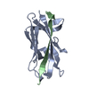
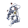
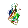
| Title | complex structure of PHF1 | ||||||
 Components Components |
| ||||||
 Keywords Keywords | GENE REGULATION / PHF1 / Tudor / histone variant / complex / Structural Genomics / Structural Genomics Consortium / SGC | ||||||
| Function / homology |  Function and homology information Function and homology informationspermatogonial cell division / histone H3K36me3 reader activity / heterochromatin organization / histone methyltransferase binding / DNA replication-dependent chromatin assembly / ESC/E(Z) complex / histone H3K4me3 reader activity / negative regulation of gene expression, epigenetic / DNA repair-dependent chromatin remodeling / regulation of cell differentiation ...spermatogonial cell division / histone H3K36me3 reader activity / heterochromatin organization / histone methyltransferase binding / DNA replication-dependent chromatin assembly / ESC/E(Z) complex / histone H3K4me3 reader activity / negative regulation of gene expression, epigenetic / DNA repair-dependent chromatin remodeling / regulation of cell differentiation / heterochromatin / Packaging Of Telomere Ends / Recognition and association of DNA glycosylase with site containing an affected purine / Cleavage of the damaged purine / Recognition and association of DNA glycosylase with site containing an affected pyrimidine / Cleavage of the damaged pyrimidine / Inhibition of DNA recombination at telomere / Meiotic synapsis / transcription corepressor binding / Condensation of Prophase Chromosomes / condensed nuclear chromosome / PRC2 methylates histones and DNA / Nonhomologous End-Joining (NHEJ) / Formation of the beta-catenin:TCF transactivating complex / G2/M DNA damage checkpoint / DNA Damage/Telomere Stress Induced Senescence / Meiotic recombination / structural constituent of chromatin / nucleosome / nucleosome assembly / Recruitment and ATM-mediated phosphorylation of repair and signaling proteins at DNA double strand breaks / site of double-strand break / chromatin organization / Processing of DNA double-strand break ends / chromosome, telomeric region / protein heterodimerization activity / chromatin binding / centrosome / DNA binding / extracellular exosome / zinc ion binding / nucleoplasm / identical protein binding / nucleus / cytosol Similarity search - Function | ||||||
| Biological species |  Homo sapiens (human) Homo sapiens (human) | ||||||
| Method |  X-RAY DIFFRACTION / SYNCHROTRON / MOLECULAR REPLACEMENT / Resolution: 1.8 Å X-RAY DIFFRACTION / SYNCHROTRON / MOLECULAR REPLACEMENT / Resolution: 1.8 Å | ||||||
 Authors Authors | Dong, C. / Bountra, C. / Edwards, A.M. / Arrowsmith, C.H. / Min, J.R. / Structural Genomics Consortium (SGC) | ||||||
 Citation Citation | Journal: Elife / Year: 2020 Title: Structural basis for histone variant H3tK27me3 recognition by PHF1 and PHF19. Authors: Dong, C. / Nakagawa, R. / Oyama, K. / Yamamoto, Y. / Zhang, W. / Dong, A. / Li, Y. / Yoshimura, Y. / Kamiya, H. / Nakayama, J.I. / Ueda, J. / Min, J. | ||||||
| History |
|
- Structure visualization
| Structure viewer | Molecule: MolmilJmol/JSmol |
|---|
- Downloads & links
-Download
| PDBx/mmCIF format | 6wat.cif.gz | 507.4 KB | Display | PDBx/mmCIF format |
|---|---|---|---|---|
| PDB format | pdb6wat.ent.gz | Display | PDB format | |
| PDBx/mmJSON format | 6wat.json.gz | Tree view | PDBx/mmJSON format | |
| Others |  Other downloads Other downloads |
-Validation report
| Arichive directory | https://data.pdbj.org/pub/pdb/validation_reports/wa/6watftp://data.pdbj.org/pub/pdb/validation_reports/wa/6wat | HTTPS FTP |
|---|
-Related structure data
| Related structure data |  6wauC  6wavC  4hczS S: Starting model for refinement C: citing same article ( |
|---|---|
| Similar structure data |


-Links
 PDBj
PDBj


- Assembly
| Deposited unit | 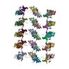
| ||||||||||||
|---|---|---|---|---|---|---|---|---|---|---|---|---|---|
| 1 | 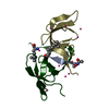
| ||||||||||||
| 2 | 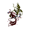
| ||||||||||||
| 3 | 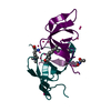
| ||||||||||||
| 4 | 
| ||||||||||||
| 5 | 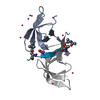
| ||||||||||||
| 6 | 
| ||||||||||||
| 7 | 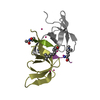
| ||||||||||||
| 8 | 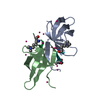
| ||||||||||||
| 9 | 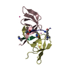
| ||||||||||||
| 10 | 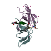
| ||||||||||||
| 11 | 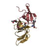
| ||||||||||||
| 12 | 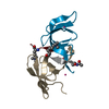
| ||||||||||||
| 13 | 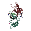
| ||||||||||||
| 14 | 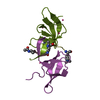
| ||||||||||||
| 15 | 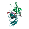
| ||||||||||||
| Unit cell |
|
-Components
| #1: Protein | Mass: 6938.849 Da / Num. of mol.: 30 Source method: isolated from a genetically manipulated source Source: (gene. exp.) Homo sapiens (human) / Gene: PHF1, PCL1 / Plasmid: pET28-MHL / Production host:  #2: Protein/peptide | Mass: 1018.231 Da / Num. of mol.: 30 / Source method: obtained synthetically / Source: (synth.) Homo sapiens (human) / References: UniProt: Q16695#3: Chemical | ChemComp-UNX /   Num. of mol.: 130 / Source method: obtained synthetically Num. of mol.: 130 / Source method: obtained synthetically#4: Water | ChemComp-HOH / |  Mass: 18.015 Da / Num. of mol.: 564 / Source method: isolated from a natural source / Formula: H2O Mass: 18.015 Da / Num. of mol.: 564 / Source method: isolated from a natural source / Formula: H2OHas ligand of interest | N | |
|---|
-Experimental details
-Experiment
| Experiment | Method: X-RAY DIFFRACTION / Number of used crystals: 1 |
|---|
- Sample preparation
| Crystal | Density Matthews: 2.43 Å3/Da / Density % sol: 49.39 % / Mosaicity: 0.14 ° |
|---|---|
| Crystal grow | Temperature: 291 K / Method: vapor diffusion, sitting drop / pH: 7 Details: 2.4M ammonium phosphate dibasic and 0.1M HEPES pH7.0 |
-Data collection
| Diffraction | Mean temperature: 100 K / Serial crystal experiment: N | ||||||||||||||||||||||||||||||
|---|---|---|---|---|---|---|---|---|---|---|---|---|---|---|---|---|---|---|---|---|---|---|---|---|---|---|---|---|---|---|---|
| Diffraction source | Source: SYNCHROTRON / Site: CLSI  / Beamline: 08ID-1 / Wavelength: 0.97625 Å / Beamline: 08ID-1 / Wavelength: 0.97625 Å | ||||||||||||||||||||||||||||||
| Detector | Type: RAYONIX MX-300 / Detector: CCD / Date: Sep 16, 2015 | ||||||||||||||||||||||||||||||
| Radiation | Protocol: SINGLE WAVELENGTH / Monochromatic (M) / Laue (L): M / Scattering type: x-ray | ||||||||||||||||||||||||||||||
| Radiation wavelength | Wavelength: 0.97625 Å / Relative weight: 1 | ||||||||||||||||||||||||||||||
| Reflection | Resolution: 1.8→47.8 Å / Num. obs: 207862 / % possible obs: 99.2 % / Redundancy: 3.8 % / CC1/2: 0.999 / Rmerge(I) obs: 0.071 / Rpim(I) all: 0.042 / Rrim(I) all: 0.082 / Net I/σ(I): 10.5 | ||||||||||||||||||||||||||||||
| Reflection shell | Diffraction-ID: 1
|
- Processing
| Software |
| |||||||||||||||||||||||||||||||||||||||||||||||||||||||||||||||||||||||||||||||||||||||||||||||||||||||||||||||||||||||||||||||||||||||||||||||||||||||||||||||||||||||||||||||||||||||||||||||||||||||||||||||||||||||||
|---|---|---|---|---|---|---|---|---|---|---|---|---|---|---|---|---|---|---|---|---|---|---|---|---|---|---|---|---|---|---|---|---|---|---|---|---|---|---|---|---|---|---|---|---|---|---|---|---|---|---|---|---|---|---|---|---|---|---|---|---|---|---|---|---|---|---|---|---|---|---|---|---|---|---|---|---|---|---|---|---|---|---|---|---|---|---|---|---|---|---|---|---|---|---|---|---|---|---|---|---|---|---|---|---|---|---|---|---|---|---|---|---|---|---|---|---|---|---|---|---|---|---|---|---|---|---|---|---|---|---|---|---|---|---|---|---|---|---|---|---|---|---|---|---|---|---|---|---|---|---|---|---|---|---|---|---|---|---|---|---|---|---|---|---|---|---|---|---|---|---|---|---|---|---|---|---|---|---|---|---|---|---|---|---|---|---|---|---|---|---|---|---|---|---|---|---|---|---|---|---|---|---|---|---|---|---|---|---|---|---|---|---|---|---|---|---|---|---|
| Refinement | Method to determine structure: MOLECULAR REPLACEMENT Starting model: 4HCZ Resolution: 1.8→40.76 Å / SU ML: 0.2387 / Cross valid method: FREE R-VALUE / σ(F): 0 / Phase error: 36.2578
| |||||||||||||||||||||||||||||||||||||||||||||||||||||||||||||||||||||||||||||||||||||||||||||||||||||||||||||||||||||||||||||||||||||||||||||||||||||||||||||||||||||||||||||||||||||||||||||||||||||||||||||||||||||||||
| Solvent computation | Shrinkage radii: 0.9 Å / VDW probe radii: 1.11 Å | |||||||||||||||||||||||||||||||||||||||||||||||||||||||||||||||||||||||||||||||||||||||||||||||||||||||||||||||||||||||||||||||||||||||||||||||||||||||||||||||||||||||||||||||||||||||||||||||||||||||||||||||||||||||||
| Displacement parameters | Biso mean: 32.19 Å2 | |||||||||||||||||||||||||||||||||||||||||||||||||||||||||||||||||||||||||||||||||||||||||||||||||||||||||||||||||||||||||||||||||||||||||||||||||||||||||||||||||||||||||||||||||||||||||||||||||||||||||||||||||||||||||
| Refinement step | Cycle: LAST / Resolution: 1.8→40.76 Å
| |||||||||||||||||||||||||||||||||||||||||||||||||||||||||||||||||||||||||||||||||||||||||||||||||||||||||||||||||||||||||||||||||||||||||||||||||||||||||||||||||||||||||||||||||||||||||||||||||||||||||||||||||||||||||
| Refine LS restraints |
| |||||||||||||||||||||||||||||||||||||||||||||||||||||||||||||||||||||||||||||||||||||||||||||||||||||||||||||||||||||||||||||||||||||||||||||||||||||||||||||||||||||||||||||||||||||||||||||||||||||||||||||||||||||||||
| LS refinement shell |
|
