 Movie
Movie Controller

[English] 日本語
 Yorodumi
- PDB-6wab: Crystal structure of human galectin-4 C-terminal carbohydrate rec... -
+ Open data

- Basic information
| Entry | Database: PDB / ID: 6wab | ||||||
|---|---|---|---|---|---|---|---|
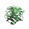
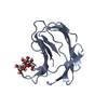
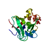
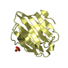
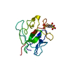
| Title | Crystal structure of human galectin-4 C-terminal carbohydrate recognition domain in complex with galactose derivative | ||||||
 Components Components | Galectin-4 | ||||||
 Keywords Keywords | SUGAR BINDING PROTEIN / GALECTIN / LECTIN / BETA SANDWICH / CARBOHYDRATE RECOGNITION / CARBOHYDRATE BINDING PROTEIN | ||||||
| Function / homology |  Function and homology information Function and homology informationantibacterial peptide biosynthetic process / galactoside binding / carbohydrate binding / extracellular matrix / cell adhesion / : / plasma membrane / cytosol Similarity search - Function | ||||||
| Biological species |  Homo sapiens (human) Homo sapiens (human) | ||||||
| Method |  X-RAY DIFFRACTION / SYNCHROTRON / MOLECULAR REPLACEMENT / Resolution: 2.28 Å X-RAY DIFFRACTION / SYNCHROTRON / MOLECULAR REPLACEMENT / Resolution: 2.28 Å | ||||||
 Authors Authors | Go, R.M. / Kishor, C. / Blanchard, H. | ||||||
 Citation Citation | Journal: To Be Published Title: Crystal structure of human galectin-4 C-terminal carbohydrate recognition domain in complex with galactose derivative Authors: Go, R.M. / Kishor, C. / Blanchard, H. | ||||||
| History |
|
- Structure visualization
| Structure viewer | Molecule: MolmilJmol/JSmol |
|---|
- Downloads & links
-Download
| PDBx/mmCIF format | 6wab.cif.gz | 126.3 KB | Display | PDBx/mmCIF format |
|---|---|---|---|---|
| PDB format | pdb6wab.ent.gz | 95.8 KB | Display | PDB format |
| PDBx/mmJSON format | 6wab.json.gz | Tree view | PDBx/mmJSON format | |
| Others |  Other downloads Other downloads |
-Validation report
| Arichive directory | https://data.pdbj.org/pub/pdb/validation_reports/wa/6wabftp://data.pdbj.org/pub/pdb/validation_reports/wa/6wab | HTTPS FTP |
|---|
-Related structure data
| Related structure data |  4ym2S S: Starting model for refinement |
|---|---|
| Similar structure data |


-Links
 PDBj
PDBj- Assembly
| Deposited unit | 
| ||||||||
|---|---|---|---|---|---|---|---|---|---|
| 1 | 
| ||||||||
| 2 | 
| ||||||||
| 3 | 
| ||||||||
| 4 | 
| ||||||||
| Unit cell |
|
-Components
| #1: Protein | Mass: 15440.567 Da / Num. of mol.: 4 / Fragment: C-terminal carbohydrate recognition domain Source method: isolated from a genetically manipulated source Source: (gene. exp.) Homo sapiens (human) / Gene: LGALS4 / Production host:  #2: Chemical |   Mass: 466.465 Da / Num. of mol.: 2 / Source method: obtained synthetically / Formula: C23H23FN6O4 / Feature type: SUBJECT OF INVESTIGATION Mass: 466.465 Da / Num. of mol.: 2 / Source method: obtained synthetically / Formula: C23H23FN6O4 / Feature type: SUBJECT OF INVESTIGATION#3: Chemical |   Mass: 92.094 Da / Num. of mol.: 2 / Source method: obtained synthetically / Formula: C3H8O3 Mass: 92.094 Da / Num. of mol.: 2 / Source method: obtained synthetically / Formula: C3H8O3#4: Water | ChemComp-HOH / |  Mass: 18.015 Da / Num. of mol.: 93 / Source method: isolated from a natural source / Formula: H2O Mass: 18.015 Da / Num. of mol.: 93 / Source method: isolated from a natural source / Formula: H2OHas ligand of interest | Y | |
|---|
-Experimental details
-Experiment
| Experiment | Method: X-RAY DIFFRACTION / Number of used crystals: 1 |
|---|
- Sample preparation
| Crystal | Density Matthews: 2.15 Å3/Da / Density % sol: 42.71 % |
|---|---|
| Crystal grow | Temperature: 293 K / Method: vapor diffusion, hanging drop / pH: 7 Details: 1.6 M ammonium sulfate, 4% (v/v) polyethylene glycol (PEG) 400, 0.1 M HEPES pH 7.0 PH range: 7 |
-Data collection
| Diffraction | Mean temperature: 100 K / Serial crystal experiment: N |
|---|---|
| Diffraction source | Source: SYNCHROTRON / Site: Australian Synchrotron  / Beamline: MX2 / Wavelength: 0.9537 Å / Beamline: MX2 / Wavelength: 0.9537 Å |
| Detector | Type: DECTRIS EIGER X 16M / Detector: PIXEL / Date: Oct 22, 2019 |
| Radiation | Protocol: SINGLE WAVELENGTH / Monochromatic (M) / Laue (L): M / Scattering type: x-ray |
| Radiation wavelength | Wavelength: 0.9537 Å / Relative weight: 1 |
| Reflection | Resolution: 2.28→45.01 Å / Num. obs: 23306 / % possible obs: 98.6 % / Redundancy: 3.4 % / CC1/2: 0.992 / Net I/σ(I): 6.8 |
| Reflection shell | Resolution: 2.28→2.36 Å / Num. unique obs: 2251 / CC1/2: 0.766 |
- Processing
| Software |
| |||||||||||||||||||||||||||||||||||||||||||||||||||||||||||||||||||||||||||||||||||||||||||||||||||||||||||||||||||||||||||||||||||||||||||||||||||||||||||
|---|---|---|---|---|---|---|---|---|---|---|---|---|---|---|---|---|---|---|---|---|---|---|---|---|---|---|---|---|---|---|---|---|---|---|---|---|---|---|---|---|---|---|---|---|---|---|---|---|---|---|---|---|---|---|---|---|---|---|---|---|---|---|---|---|---|---|---|---|---|---|---|---|---|---|---|---|---|---|---|---|---|---|---|---|---|---|---|---|---|---|---|---|---|---|---|---|---|---|---|---|---|---|---|---|---|---|---|---|---|---|---|---|---|---|---|---|---|---|---|---|---|---|---|---|---|---|---|---|---|---|---|---|---|---|---|---|---|---|---|---|---|---|---|---|---|---|---|---|---|---|---|---|---|---|---|---|
| Refinement | Method to determine structure: MOLECULAR REPLACEMENT Starting model: 4YM2 Resolution: 2.28→36.795 Å / Cor.coef. Fo:Fc: 0.945 / Cor.coef. Fo:Fc free: 0.907 / SU B: 8.744 / SU ML: 0.216 / Cross valid method: FREE R-VALUE / ESU R: 0.49 / ESU R Free: 0.281 Details: Hydrogens have been added in their riding positions
| |||||||||||||||||||||||||||||||||||||||||||||||||||||||||||||||||||||||||||||||||||||||||||||||||||||||||||||||||||||||||||||||||||||||||||||||||||||||||||
| Solvent computation | Ion probe radii: 0.8 Å / Shrinkage radii: 0.8 Å / VDW probe radii: 1.2 Å | |||||||||||||||||||||||||||||||||||||||||||||||||||||||||||||||||||||||||||||||||||||||||||||||||||||||||||||||||||||||||||||||||||||||||||||||||||||||||||
| Displacement parameters | Biso mean: 36.074 Å2
| |||||||||||||||||||||||||||||||||||||||||||||||||||||||||||||||||||||||||||||||||||||||||||||||||||||||||||||||||||||||||||||||||||||||||||||||||||||||||||
| Refinement step | Cycle: LAST / Resolution: 2.28→36.795 Å
| |||||||||||||||||||||||||||||||||||||||||||||||||||||||||||||||||||||||||||||||||||||||||||||||||||||||||||||||||||||||||||||||||||||||||||||||||||||||||||
| Refine LS restraints |
| |||||||||||||||||||||||||||||||||||||||||||||||||||||||||||||||||||||||||||||||||||||||||||||||||||||||||||||||||||||||||||||||||||||||||||||||||||||||||||
| LS refinement shell |
|
