[English] 日本語
Yorodumi
- PDB-6w2i: Crystal Structure of Y188G Variant of the Internal UBA Domain of ... -

+
Open data


ID or keywords:

Loading...

-
Basic information

Entry
Database: PDB / ID: 6w2i
Title
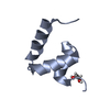
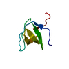
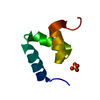
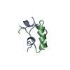
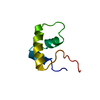
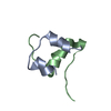
Crystal Structure of Y188G Variant of the Internal UBA Domain of HHR23A
ComponentsUV excision repair protein RAD23 homolog A
KeywordsDNA BINDING PROTEIN / Ubiquitin Associated Domain / UBA Domain / Helical Bundle
Function / homology
Function and homology information


regulation of proteasomal ubiquitin-dependent protein catabolic process / proteasome binding / ubiquitin-specific protease binding / polyubiquitin modification-dependent protein binding / positive regulation of viral genome replication / positive regulation of cell cycle / proteasome complex / Josephin domain DUBs / ubiquitin binding / nucleotide-excision repair ...regulation of proteasomal ubiquitin-dependent protein catabolic process / proteasome binding / ubiquitin-specific protease binding / polyubiquitin modification-dependent protein binding / positive regulation of viral genome replication / positive regulation of cell cycle / proteasome complex / Josephin domain DUBs / ubiquitin binding / nucleotide-excision repair / DNA Damage Recognition in GG-NER / protein destabilization / kinase binding / Formation of Incision Complex in GG-NER / positive regulation of proteasomal ubiquitin-dependent protein catabolic process / single-stranded DNA binding / proteasome-mediated ubiquitin-dependent protein catabolic process / damaged DNA binding / intracellular membrane-bounded organelle / Golgi apparatus / protein-containing complex / nucleoplasm / nucleus / cytoplasm / cytosol
Similarity search - Function
RAD23A/RAD23B, UBA1 domain / UV excision repair protein Rad23 / XPC-binding domain / XPC-binding domain superfamily / XPC-binding domain / Heat shock chaperonin-binding / Heat shock chaperonin-binding motif. / UBA/TS-N domain / Ubiquitin associated domain / Ubiquitin-associated domain ...RAD23A/RAD23B, UBA1 domain / UV excision repair protein Rad23 / XPC-binding domain / XPC-binding domain superfamily / XPC-binding domain / Heat shock chaperonin-binding / Heat shock chaperonin-binding motif. / UBA/TS-N domain / Ubiquitin associated domain / Ubiquitin-associated domain / Ubiquitin-associated domain (UBA) profile. / UBA-like superfamily / Ubiquitin family / Ubiquitin homologues / Ubiquitin domain profile. / Ubiquitin-like domain / Ubiquitin-like domain superfamily
Similarity search - Domain/homology
UV excision repair protein RAD23 homolog A
Similarity search - Component
Biological speciesHomo sapiens (human)
MethodX-RAY DIFFRACTION / SYNCHROTRON / SAD / Resolution: 1.45 Å
AuthorsBowler, B.E. / Zeng, B. / Becht, D.C. / Rothfuss, M. / Sprang, S.R. / Mou, T.-C.
Funding support United States, 2items
OrganizationGrant numberCountry
National Science Foundation (NSF, United States)MCB-1412164 United States
National Institutes of Health/National Institute of General Medical Sciences (NIH/NIGMS)P20GM103546 United States
CitationJournal: J.Am.Chem.Soc. / Year: 2023
Title: High-Accuracy Prediction of Stabilizing Surface Mutations to the Three-Helix Bundle, UBA(1), with EmCAST.
Authors: Rothfuss, M.T. / Becht, D.C. / Zeng, B. / McClelland, L.J. / Yates-Hansen, C. / Bowler, B.E.
History
DepositionMar 5, 2020Deposition site: RCSB / Processing site: RCSB
Revision 1.0Mar 10, 2021Provider: repository / Type: Initial release
Revision 2.0Aug 25, 2021Group: Advisory / Atomic model ...Advisory / Atomic model / Data collection / Database references / Derived calculations / Structure summary
Category: atom_site / database_2 ...atom_site / database_2 / pdbx_nonpoly_scheme / pdbx_poly_seq_scheme / pdbx_unobs_or_zero_occ_atoms / pdbx_unobs_or_zero_occ_residues / struct_conf / struct_ref_seq / struct_ref_seq_dif / struct_site / struct_site_gen
Item: _atom_site.B_iso_or_equiv / _atom_site.Cartn_x ..._atom_site.B_iso_or_equiv / _atom_site.Cartn_x / _atom_site.Cartn_y / _atom_site.Cartn_z / _atom_site.auth_seq_id / _database_2.pdbx_DOI / _database_2.pdbx_database_accession / _pdbx_nonpoly_scheme.auth_seq_num / _pdbx_nonpoly_scheme.pdb_seq_num / _pdbx_poly_seq_scheme.pdb_seq_num / _pdbx_unobs_or_zero_occ_residues.auth_seq_id / _struct_conf.beg_auth_seq_id / _struct_conf.end_auth_seq_id / _struct_ref_seq.pdbx_auth_seq_align_beg / _struct_ref_seq.pdbx_auth_seq_align_end / _struct_ref_seq_dif.pdbx_auth_seq_num / _struct_site.details / _struct_site.pdbx_auth_seq_id / _struct_site_gen.auth_seq_id
Revision 2.1Nov 1, 2023Group: Data collection / Database references
Category: chem_comp_atom / chem_comp_bond ...chem_comp_atom / chem_comp_bond / citation / citation_author
Item: _citation.country / _citation.journal_abbrev ..._citation.country / _citation.journal_abbrev / _citation.journal_id_ASTM / _citation.journal_id_CSD / _citation.journal_id_ISSN / _citation.journal_volume / _citation.page_first / _citation.page_last / _citation.pdbx_database_id_DOI / _citation.pdbx_database_id_PubMed / _citation.title / _citation.year

-
Structure visualization

Structure viewerMolecule:
MolmilJmol/JSmol

Downloads & links

-
Assembly

Deposited unit
A: UV excision repair protein RAD23 homolog A
hetero molecules


Theoretical massNumber of molelcules
Total (without water)5,6262
Polymers5,5341
Non-polymers921
Water86548
1


  • Idetical with deposited unit
  • defined by author&software
TypeNameSymmetry operationNumber
identity operation1_555x,y,z1
MethodPISA
Unit cell
Length a, b, c (Å)31.164, 31.164, 40.530
Angle α, β, γ (deg.)90.000, 90.000, 90.000
Int Tables number78
Space group name H-MP43
Space group name HallP4cw
Symmetry operation#1: x,y,z
#2: -y,x,z+3/4
#3: y,-x,z+1/4
#4: -x,-y,z+1/2

-
Components

#1: Protein UV excision repair protein RAD23 homolog A / hHR23A


Mass: 5534.243 Da / Num. of mol.: 1 / Mutation: Y188G
Source method: isolated from a genetically manipulated source
Source: (gene. exp.) Homo sapiens (human) / Gene: RAD23A / Plasmid: pGEX-2T
Details (production host): Thrombin cleavage site replaced with TEV protease cleavage site
Production host: Escherichia coli BL21(DE3) (bacteria) / Strain (production host): BL21(DE3) / References: UniProt: P54725
#2: Chemical ChemComp-GOL / GLYCEROL / GLYCERIN / PROPANE-1,2,3-TRIOL


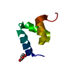
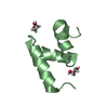
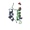
Mass: 92.094 Da / Num. of mol.: 1 / Source method: obtained synthetically / Formula: C3H8O3
#3: Water ChemComp-HOH / water


Mass: 18.015 Da / Num. of mol.: 48 / Source method: isolated from a natural source / Formula: H2O
Has ligand of interestN

-
Experimental details

-
Experiment

ExperimentMethod: X-RAY DIFFRACTION / Number of used crystals: 2

-
Sample preparation

Crystal
IDDensity Matthews3/Da)Density % sol (%)
11.7830.83
2
Crystal grow
Temperature (K)Crystal-IDMethodDetails
2931vapor diffusion, sitting drop2.0M Ammonium sulfate
2932vapor diffusion, sitting drop2.0M Ammonium sulfate

-
Data collection

Diffraction
IDMean temperature (K)Crystal-IDSerial crystal experiment
11001N
21002N
Diffraction source
SourceSiteBeamlineIDWavelength (Å)
SYNCHROTRONSSRL BL9-210.9795
SYNCHROTRONSSRL BL9-221.77
Detector
TypeIDDetectorDate
DECTRIS PILATUS 6M1PIXELJan 29, 2020
DECTRIS PILATUS 6M2PIXELJan 29, 2020
Radiation
IDProtocolMonochromatic (M) / Laue (L)Scattering typeWavelength-ID
1SINGLE WAVELENGTHMx-ray1
2SINGLE WAVELENGTHMx-ray2
Radiation wavelength
IDWavelength (Å)Relative weight
10.97951
21.771
Reflection

Entry-ID: 6W2I / CC1/2: 1 / CC star: 1

Resolution (Å)Num. obs% possible obs (%)Redundancy (%)Biso Wilson estimate2)Rmerge(I) obsRpim(I) allRrim(I) allDiffraction-IDNet I/σ(I)
1.45-31.16687598.9513.411.230.076580.021530.07961122.37
1.9-31.14308399.143.516.480.037590.0055480.03801293.75
Reflection shell
Resolution (Å)Redundancy (%)Rmerge(I) obsMean I/σ(I) obsNum. unique obsCC1/2CC starRpim(I) allRrim(I) allDiffraction-ID% possible all
1.45-1.50212.50.59014.346800.9260.980.17030.615197.84
1.9-1.96824.10.0913529.742920.99910.01890.09335296.05

-
Processing

Software
NameVersionClassification
Blu-Icedata collection
XDSMar 15, 2019data reduction
Aimless0.7.4data scaling
HKL2Map0.4.e-betaphasing
AutoSolphasing
PHENIX1.17.1-3660model building
Coot0.8.9.2 ELmodel building
PHENIX1.17.1-3660refinement
RefinementMethod to determine structure: SAD / Resolution: 1.45→31.16 Å / SU ML: 0.1067 / Cross valid method: FREE R-VALUE / σ(F): 1.36 / Phase error: 19.1748
RfactorNum. reflection% reflection
Rfree0.183 697 10.14 %
Rwork0.1498 --
obs0.1532 6875 98.96 %
Solvent computationShrinkage radii: 0.9 Å / VDW probe radii: 1.11 Å
Displacement parametersBiso mean: 15.14 Å2
Refinement stepCycle: LAST / Resolution: 1.45→31.16 Å
ProteinNucleic acidLigandSolventTotal
Num. atoms369 0 6 48 423
Refine LS restraints
Refine-IDTypeDev idealNumber
X-RAY DIFFRACTIONf_bond_d0.0112388
X-RAY DIFFRACTIONf_angle_d1.0683524
X-RAY DIFFRACTIONf_chiral_restr0.090459
X-RAY DIFFRACTIONf_plane_restr0.006868
X-RAY DIFFRACTIONf_dihedral_angle_d6.32860
LS refinement shell
Resolution (Å)Rfactor RfreeNum. reflection RfreeRfactor RworkNum. reflection RworkRefine-ID% reflection obs (%)
1.45-1.560.19391340.15131223X-RAY DIFFRACTION98.19
1.56-1.720.1921400.14351222X-RAY DIFFRACTION99.05
1.72-1.970.19371400.14271229X-RAY DIFFRACTION98.92
1.97-2.480.17741430.14391248X-RAY DIFFRACTION99.78
2.48-31.160.17871400.15651256X-RAY DIFFRACTION98.87

+
About Yorodumi

-
News

-
Feb 9, 2022. New format data for meta-information of EMDB entries

New format data for meta-information of EMDB entries

  • Version 3 of the EMDB header file is now the official format.
  • The previous official version 1.9 will be removed from the archive.

Related info.:EMDB header

External links:wwPDB to switch to version 3 of the EMDB data model

-
Aug 12, 2020. Covid-19 info

Covid-19 info

URL: https://pdbjlvh1.pdbj.org/emnavi/covid19.php

New page: Covid-19 featured information page in EM Navigator.

Related info.:Covid-19 info / Mar 5, 2020. Novel coronavirus structure data

+
Mar 5, 2020. Novel coronavirus structure data

Novel coronavirus structure data

Related info.:Yorodumi Speices / Aug 12, 2020. Covid-19 info

External links:COVID-19 featured content - PDBj / Molecule of the Month (242):Coronavirus Proteases

+
Jan 31, 2019. EMDB accession codes are about to change! (news from PDBe EMDB page)

EMDB accession codes are about to change! (news from PDBe EMDB page)

  • The allocation of 4 digits for EMDB accession codes will soon come to an end. Whilst these codes will remain in use, new EMDB accession codes will include an additional digit and will expand incrementally as the available range of codes is exhausted. The current 4-digit format prefixed with “EMD-” (i.e. EMD-XXXX) will advance to a 5-digit format (i.e. EMD-XXXXX), and so on. It is currently estimated that the 4-digit codes will be depleted around Spring 2019, at which point the 5-digit format will come into force.
  • The EM Navigator/Yorodumi systems omit the EMD- prefix.

Related info.:Q: What is EMD? / ID/Accession-code notation in Yorodumi/EM Navigator

External links:EMDB Accession Codes are Changing Soon! / Contact to PDBj

+
Jul 12, 2017. Major update of PDB

Major update of PDB

  • wwPDB released updated PDB data conforming to the new PDBx/mmCIF dictionary.
  • This is a major update changing the version number from 4 to 5, and with Remediation, in which all the entries are updated.
  • In this update, many items about electron microscopy experimental information are reorganized (e.g. em_software).
  • Now, EM Navigator and Yorodumi are based on the updated data.

External links:wwPDB Remediation / Enriched Model Files Conforming to OneDep Data Standards Now Available in the PDB FTP Archive

-
Yorodumi

Thousand views of thousand structures

  • Yorodumi is a browser for structure data from EMDB, PDB, SASBDB, etc.
  • This page is also the successor to EM Navigator detail page, and also detail information page/front-end page for Omokage search.
  • The word "yorodu" (or yorozu) is an old Japanese word meaning "ten thousand". "mi" (miru) is to see.

Related info.:EMDB / PDB / SASBDB / Comparison of 3 databanks / Yorodumi Search / Aug 31, 2016. New EM Navigator & Yorodumi / Yorodumi Papers / Jmol/JSmol / Function and homology information / Changes in new EM Navigator and Yorodumi

Read more