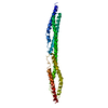
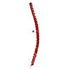
- PDB-6vzf: Crystal Structure of Atg11 Coiled-Coil 3 -
+
Open data
ID or keywords:
Loading...
-
Basic information
Entry
Database: PDB / ID: 6vzf
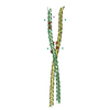
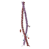
Title
Crystal Structure of Atg11 Coiled-Coil 3
Components
Autophagy-related protein 11
Keywords
STRUCTURAL PROTEIN / coiled-coil / autophagy / scaffold
Function / homology
Function and homology information
autophagy of peroxisome / Macroautophagy / Atg1/ULK1 kinase complex / cytoplasm to vacuole targeting by the Cvt pathway / positive regulation of autophagosome assembly / ribophagy / phagophore assembly site membrane / protein localization to phagophore assembly site / autophagy of mitochondrion / piecemeal microautophagy of the nucleus ...autophagy of peroxisome / Macroautophagy / Atg1/ULK1 kinase complex / cytoplasm to vacuole targeting by the Cvt pathway / positive regulation of autophagosome assembly / ribophagy / phagophore assembly site membrane / protein localization to phagophore assembly site / autophagy of mitochondrion / piecemeal microautophagy of the nucleus / pexophagy / phagophore assembly site / reticulophagy / cellular response to nitrogen starvation / protein-containing complex localization / vacuolar membrane / autophagosome assembly / SNARE binding / positive regulation of protein phosphorylation / autophagy / molecular adaptor activity / protein kinase binding Similarity search - Function
Autophagy protein ATG17-like domain / Autophagy protein ATG17-like domain / Autophagy-related protein 11, C-terminal / Autophagy-related protein 11 / Autophagy-related protein 11 Similarity search - Domain/homology
Method to determine structure: SAD / Resolution: 2.03→34.61 Å / Cor.coef. Fo:Fc: 0.946 / Cor.coef. Fo:Fc free: 0.933 / SU B: 2.842 / SU ML: 0.078 / SU R Cruickshank DPI: 0.119 / Cross valid method: THROUGHOUT / σ(F): 0 / ESU R: 0.119 / ESU R Free: 0.115 Details: HYDROGENS HAVE BEEN ADDED IN THE RIDING POSITIONS U VALUES : REFINED INDIVIDUALLY
Rfactor
Num. reflection
% reflection
Selection details
Rfree
0.2459
781
5.2 %
RANDOM
Rwork
0.2263
-
-
-
obs
0.2273
14270
99.93 %
-
Solvent computation
Ion probe radii: 0.8 Å / Shrinkage radii: 0.8 Å / VDW probe radii: 1.2 Å
In the structure databanks used in Yorodumi, some data are registered as the other names, "COVID-19 virus" and "2019-nCoV". Here are the details of the virus and the list of structure data.
Jan 31, 2019. EMDB accession codes are about to change! (news from PDBe EMDB page)
EMDB accession codes are about to change! (news from PDBe EMDB page)
The allocation of 4 digits for EMDB accession codes will soon come to an end. Whilst these codes will remain in use, new EMDB accession codes will include an additional digit and will expand incrementally as the available range of codes is exhausted. The current 4-digit format prefixed with “EMD-” (i.e. EMD-XXXX) will advance to a 5-digit format (i.e. EMD-XXXXX), and so on. It is currently estimated that the 4-digit codes will be depleted around Spring 2019, at which point the 5-digit format will come into force.
The EM Navigator/Yorodumi systems omit the EMD- prefix.
Related info.:Q: What is EMD? / ID/Accession-code notation in Yorodumi/EM Navigator
Yorodumi is a browser for structure data from EMDB, PDB, SASBDB, etc.
This page is also the successor to EM Navigator detail page, and also detail information page/front-end page for Omokage search.
The word "yorodu" (or yorozu) is an old Japanese word meaning "ten thousand". "mi" (miru) is to see.
Related info.:EMDB / PDB / SASBDB / Comparison of 3 databanks / Yorodumi Search / Aug 31, 2016. New EM Navigator & Yorodumi / Yorodumi Papers / Jmol/JSmol / Function and homology information / Changes in new EM Navigator and Yorodumi
 Movie
Movie Controller


 Open data
 Basic information
 Components
 Keywords
 Function and homology information
 X-RAY DIFFRACTION /
  Authors
 United States, 2items
  Citation
 Structure visualization
 Downloads & links
 Other downloads


 PDBj
PDBj
 Assembly
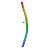


 Mass: 96.063 Da / Num. of mol.: 1 / Source method: obtained synthetically / Formula: SO4
 Mass: 18.015 Da / Num. of mol.: 23 / Source method: isolated from a natural source / Formula: H2O
 Sample preparation
 Processing