 Movie
Movie Controller

[English] 日本語
 Yorodumi
- PDB-6vva: N-Acetylmannosamine-6-phosphate 2-epimerase from Staphylococcus a... -
+ Open data

- Basic information
| Entry | Database: PDB / ID: 6vva | |||||||||
|---|---|---|---|---|---|---|---|---|---|---|
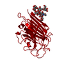
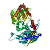
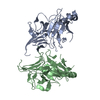
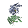
| Title | N-Acetylmannosamine-6-phosphate 2-epimerase from Staphylococcus aureus (strain MRSA USA300) | |||||||||
 Components Components | N-acetylmannosamine-6-phosphate 2-epimerase | |||||||||
 Keywords Keywords | ISOMERASE / NanE / TIM-barrel / epimerase | |||||||||
| Function / homology |  Function and homology information Function and homology information: / N-acylglucosamine-6-phosphate 2-epimerase / N-acetylmannosamine catabolic process / N-acylglucosamine-6-phosphate 2-epimerase activity / N-acetylneuraminate catabolic process / carbohydrate metabolic process / cytosol Similarity search - Function | |||||||||
| Biological species |   Staphylococcus aureus (bacteria) Staphylococcus aureus (bacteria) | |||||||||
| Method |  X-RAY DIFFRACTION / SYNCHROTRON / MOLECULAR REPLACEMENT / molecular replacement / Resolution: 1.84 Å X-RAY DIFFRACTION / SYNCHROTRON / MOLECULAR REPLACEMENT / molecular replacement / Resolution: 1.84 Å | |||||||||
 Authors Authors | Renwick, R.C.J. / Currie, M.J. | |||||||||
 Citation Citation | Journal: J.Biol.Chem. / Year: 2021 Title: N-acetylmannosamine-6-phosphate 2-epimerase uses a novel substrate-assisted mechanism to catalyze amino sugar epimerization. Authors: Currie, M.J. / Manjunath, L. / Horne, C.R. / Rendle, P.M. / Subramanian, R. / Friemann, R. / Fairbanks, A.J. / Muscroft-Taylor, A.C. / North, R.A. / Dobson, R.C.J. | |||||||||
| History |
|
- Structure visualization
| Structure viewer | Molecule: MolmilJmol/JSmol |
|---|
- Downloads & links
-Download
| PDBx/mmCIF format | 6vva.cif.gz | 117.8 KB | Display | PDBx/mmCIF format |
|---|---|---|---|---|
| PDB format | pdb6vva.ent.gz | 89.2 KB | Display | PDB format |
| PDBx/mmJSON format | 6vva.json.gz | Tree view | PDBx/mmJSON format | |
| Others |  Other downloads Other downloads |
-Validation report
| Summary document | 6vva_validation.pdf.gz | 312 KB | Display | wwPDB validaton report |
|---|---|---|---|---|
| Full document | 6vva_full_validation.pdf.gz | 312 KB | Display | |
| Data in XML | 6vva_validation.xml.gz | 1.3 KB | Display | |
| Data in CIF | 6vva_validation.cif.gz | 9.2 KB | Display | |
| Arichive directory | https://data.pdbj.org/pub/pdb/validation_reports/vv/6vvaftp://data.pdbj.org/pub/pdb/validation_reports/vv/6vva | HTTPS FTP |
-Related structure data
| Related structure data |  7mfnC  7mfsC  7mqtC  1y0eS S: Starting model for refinement C: citing same article ( |
|---|---|
| Similar structure data |


-Links
 PDBj
PDBj
- Assembly
| Deposited unit | 
| ||||||||
|---|---|---|---|---|---|---|---|---|---|
| 1 | 
| ||||||||
| Unit cell |
|
-Components
| #1: Protein | Mass: 24571.072 Da / Num. of mol.: 2 Source method: isolated from a genetically manipulated source Source: (gene. exp.) Staphylococcus aureus (bacteria)Gene: nanE, BTN44_01590, EP54_02960, EQ90_08785, ERS072840_01491, FA040_00085, HMPREF3211_02501, NCTC10654_00351, NCTC10702_00560, NCTC7878_00328, RK64_02155 Production host: References: UniProt: X5EM89, UniProt: Q2G157*PLUS, N-acylglucosamine-6-phosphate 2-epimerase #2: Chemical |   Mass: 192.124 Da / Num. of mol.: 2 / Source method: obtained synthetically / Formula: C6H8O7 / Feature type: SUBJECT OF INVESTIGATION Mass: 192.124 Da / Num. of mol.: 2 / Source method: obtained synthetically / Formula: C6H8O7 / Feature type: SUBJECT OF INVESTIGATION#3: Chemical |   Mass: 35.453 Da / Num. of mol.: 2 / Source method: obtained synthetically / Formula: Cl Mass: 35.453 Da / Num. of mol.: 2 / Source method: obtained synthetically / Formula: Cl#4: Water | ChemComp-HOH / |  Mass: 18.015 Da / Num. of mol.: 585 / Source method: isolated from a natural source / Formula: H2O Mass: 18.015 Da / Num. of mol.: 585 / Source method: isolated from a natural source / Formula: H2OHas ligand of interest | Y | |
|---|
-Experimental details
-Experiment
| Experiment | Method: X-RAY DIFFRACTION / Number of used crystals: 1 |
|---|
- Sample preparation
| Crystal | Density Matthews: 3.03 Å3/Da / Density % sol: 59.36 % |
|---|---|
| Crystal grow | Temperature: 293.15 K / Method: vapor diffusion, sitting drop Details: 1 M trisodium citrate, 0.1 M sodium cacodylate, pH 6.5 |
-Data collection
| Diffraction | Mean temperature: 100 K / Serial crystal experiment: N |
|---|---|
| Diffraction source | Source: SYNCHROTRON / Site: Australian Synchrotron  / Beamline: MX2 / Wavelength: 0.9537 Å / Beamline: MX2 / Wavelength: 0.9537 Å |
| Detector | Type: ADSC QUANTUM 315r / Detector: CCD / Date: Jul 14, 2013 |
| Radiation | Protocol: SINGLE WAVELENGTH / Monochromatic (M) / Laue (L): M / Scattering type: x-ray |
| Radiation wavelength | Wavelength: 0.9537 Å / Relative weight: 1 |
| Reflection | Resolution: 1.84→46.34 Å / Num. obs: 52642 / % possible obs: 99.7 % / Redundancy: 3.6 % / CC1/2: 0.995 / Net I/σ(I): 10.8 |
| Reflection shell | Resolution: 1.84→1.906 Å / Num. unique obs: 5094 / CC1/2: 0.592 |
-Phasing
| Phasing | Method: molecular replacement |
|---|
- Processing
| Software |
| ||||||||||||||||||||||||||||||||||||||||||||||||||||||||||||
|---|---|---|---|---|---|---|---|---|---|---|---|---|---|---|---|---|---|---|---|---|---|---|---|---|---|---|---|---|---|---|---|---|---|---|---|---|---|---|---|---|---|---|---|---|---|---|---|---|---|---|---|---|---|---|---|---|---|---|---|---|---|
| Refinement | Method to determine structure: MOLECULAR REPLACEMENT Starting model: 1Y0E Resolution: 1.84→46.34 Å / Cor.coef. Fo:Fc: 0.953 / Cor.coef. Fo:Fc free: 0.94 / SU B: 3.021 / SU ML: 0.087 / Cross valid method: THROUGHOUT / σ(F): 0 / ESU R: 0.12 / ESU R Free: 0.109 / Stereochemistry target values: MAXIMUM LIKELIHOOD Details: HYDROGENS HAVE BEEN ADDED IN THE RIDING POSITIONS U VALUES : REFINED INDIVIDUALLY
| ||||||||||||||||||||||||||||||||||||||||||||||||||||||||||||
| Solvent computation | Ion probe radii: 0.8 Å / Shrinkage radii: 0.8 Å / VDW probe radii: 1.2 Å / Solvent model: MASK | ||||||||||||||||||||||||||||||||||||||||||||||||||||||||||||
| Displacement parameters | Biso max: 54.6 Å2 / Biso mean: 16.312 Å2 / Biso min: 9.45 Å2
| ||||||||||||||||||||||||||||||||||||||||||||||||||||||||||||
| Refinement step | Cycle: final / Resolution: 1.84→46.34 Å
| ||||||||||||||||||||||||||||||||||||||||||||||||||||||||||||
| Refine LS restraints |
| ||||||||||||||||||||||||||||||||||||||||||||||||||||||||||||
| LS refinement shell | Resolution: 1.84→1.888 Å / Rfactor Rfree error: 0 / Total num. of bins used: 20
|
