 Movie
Movie Controller

[English] 日本語
 Yorodumi
- PDB-4i2n: Crystal structure of 31kD Heat Shock Protein, VcHsp31 from Vibrio... -
+ Open data

- Basic information
| Entry | Database: PDB / ID: 4i2n | ||||||
|---|---|---|---|---|---|---|---|
| Title | Crystal structure of 31kD Heat Shock Protein, VcHsp31 from Vibrio cholerae | ||||||
 Components Components | Intracellular protease/amidase | ||||||
 Keywords Keywords | HYDROLASE / Hsp-31 / Chaperone-protease / alpha-beta domains / Heat Shock Protein / small peptides and denatured proteins | ||||||
| Function / homology |  Function and homology information Function and homology informationglyoxalase III activity / : / protein deglycase activity / DNA repair / cytoplasm Similarity search - Function | ||||||
| Biological species |   Vibrio cholerae (bacteria) Vibrio cholerae (bacteria) | ||||||
| Method |  X-RAY DIFFRACTION / MOLECULAR REPLACEMENT / Resolution: 1.9 Å X-RAY DIFFRACTION / MOLECULAR REPLACEMENT / Resolution: 1.9 Å | ||||||
 Authors Authors | Das, S. / Sen, U. | ||||||
 Citation Citation | Journal: To be Published Title: Temperature dependent structural flexibility and functional activation of 31kD Heat Shock Protein, VcHsp31 from Vibrio cholerae Authors: Das, S. / Sen, U. | ||||||
| History |
|
- Structure visualization
| Structure viewer | Molecule: MolmilJmol/JSmol |
|---|
- Downloads & links
-Download
| PDBx/mmCIF format | 4i2n.cif.gz | 364.6 KB | Display | PDBx/mmCIF format |
|---|---|---|---|---|
| PDB format | pdb4i2n.ent.gz | 296.3 KB | Display | PDB format |
| PDBx/mmJSON format | 4i2n.json.gz | Tree view | PDBx/mmJSON format | |
| Others |  Other downloads Other downloads |
-Validation report
| Arichive directory | https://data.pdbj.org/pub/pdb/validation_reports/i2/4i2nftp://data.pdbj.org/pub/pdb/validation_reports/i2/4i2n | HTTPS FTP |
|---|
-Related structure data


-Links
 PDBj
PDBj

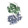
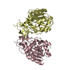
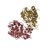
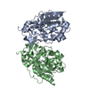
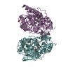
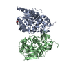
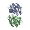
- Assembly
| Deposited unit | 
| ||||||||
|---|---|---|---|---|---|---|---|---|---|
| 1 | 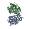
| ||||||||
| 2 | 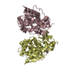
| ||||||||
| 3 | 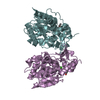
| ||||||||
| Unit cell |
|
-Components
| #1: Protein | Mass: 31367.426 Da / Num. of mol.: 6 / Fragment: UNP residues 6-284 Source method: isolated from a genetically manipulated source Source: (gene. exp.) Vibrio cholerae (bacteria) / Strain: ATCC 39541 / Ogawa 395 / O395 / Gene: A5F0Q0, VC0395_0351, VC395_A0912 / Production host: #2: Chemical | ChemComp-MPD / (   Mass: 118.174 Da / Num. of mol.: 5 / Source method: obtained synthetically / Formula: C6H14O2 / Comment: precipitant*YM Mass: 118.174 Da / Num. of mol.: 5 / Source method: obtained synthetically / Formula: C6H14O2 / Comment: precipitant*YM#3: Chemical | ChemComp-CA /   Mass: 40.078 Da / Num. of mol.: 6 / Source method: obtained synthetically / Formula: Ca Mass: 40.078 Da / Num. of mol.: 6 / Source method: obtained synthetically / Formula: Ca#4: Water | ChemComp-HOH / |  Mass: 18.015 Da / Num. of mol.: 1776 / Source method: isolated from a natural source / Formula: H2O Mass: 18.015 Da / Num. of mol.: 1776 / Source method: isolated from a natural source / Formula: H2O |
|---|
-Experimental details
-Experiment
| Experiment | Method: X-RAY DIFFRACTION / Number of used crystals: 1 |
|---|
- Sample preparation
| Crystal | Density Matthews: 2.05 Å3/Da / Density % sol: 39.94 % |
|---|---|
| Crystal grow | Temperature: 293 K / Method: vapor diffusion / pH: 5 Details: 5% PEG6000, 8% MPD, pH 5.0, VAPOR DIFFUSION, temperature 293K |
-Data collection
| Diffraction | Mean temperature: 100 K |
|---|---|
| Diffraction source | Source: ROTATING ANODE / Type: RIGAKU RU200 / Wavelength: 1.54 Å |
| Detector | Type: MAR scanner 345 mm plate / Detector: IMAGE PLATE / Date: Jun 20, 2011 |
| Radiation | Monochromator: Cu K[alpha] / Protocol: SINGLE WAVELENGTH / Monochromatic (M) / Laue (L): M / Scattering type: x-ray |
| Radiation wavelength | Wavelength: 1.54 Å / Relative weight: 1 |
| Reflection | Resolution: 1.9→30 Å / Num. all: 206188 / Num. obs: 113580 / % possible obs: 98.1 % / Observed criterion σ(F): 0 / Observed criterion σ(I): 0 / Redundancy: 1.79 % / Biso Wilson estimate: 20.4 Å2 |
| Reflection shell | Resolution: 1.9→1.99 Å / Rmerge(I) obs: 0.28 / Mean I/σ(I) obs: 2 / % possible all: 91 |
- Processing
| Software |
| ||||||||||||||||||||||||||||||||||||
|---|---|---|---|---|---|---|---|---|---|---|---|---|---|---|---|---|---|---|---|---|---|---|---|---|---|---|---|---|---|---|---|---|---|---|---|---|---|
| Refinement | Method to determine structure: MOLECULAR REPLACEMENT / Resolution: 1.9→29.54 Å / Rfactor Rfree error: 0.003 / Data cutoff high absF: 1438620.92 / Data cutoff low absF: 0 / Isotropic thermal model: RESTRAINED / Cross valid method: THROUGHOUT / σ(F): 0 / Stereochemistry target values: Engh & Huber / Details: BULK SOLVENT MODEL USED
| ||||||||||||||||||||||||||||||||||||
| Solvent computation | Solvent model: FLAT MODEL / Bsol: 58.0033 Å2 / ksol: 0.4 e/Å3 | ||||||||||||||||||||||||||||||||||||
| Displacement parameters | Biso mean: 24.6 Å2
| ||||||||||||||||||||||||||||||||||||
| Refine analyze |
| ||||||||||||||||||||||||||||||||||||
| Refinement step | Cycle: LAST / Resolution: 1.9→29.54 Å
| ||||||||||||||||||||||||||||||||||||
| Refine LS restraints |
| ||||||||||||||||||||||||||||||||||||
| Refine LS restraints NCS | NCS model details: NONE | ||||||||||||||||||||||||||||||||||||
| LS refinement shell | Resolution: 1.9→1.99 Å / Rfactor Rfree error: 0.01 / Total num. of bins used: 8
| ||||||||||||||||||||||||||||||||||||
| Xplor file |
|
